Medical term:
ECHO
ECHO
Abbreviation for enteric cytopathic human orphan. See: ECHO virus.
ech·o
(ek'ō),1. A reverberating sound sometimes heard during auscultation of the chest.
2. In ultrasonography, the acoustic signal received from scattering or reflecting structures or the corresponding pattern of light on a CRT or ultrasonogram.
3. In magnetic resonance imaging, the signal detected following an inverting pulse.
[G.]
Farlex Partner Medical Dictionary © Farlex 2012
ech·o
(ek'ō)1. A reverberating sound sometimes heard during auscultation of the chest.
2. ultrasonography The acoustic signal received from scattering or reflecting structures, or the corresponding pattern of light on a CRT or ultrasonogram.
3. magnetic resonance imaging The signal detected following an inverting pulse.
[G.]
Medical Dictionary for the Health Professions and Nursing © Farlex 2012
ultrasonography
A technique utilizing high frequency ultrasound waves (greater than 18 000 Hz) emitted by a transducer placed near the eye. The silicone probe, which rests on the eye, is separated from the transducer by a water column to segregate the noise from the transducer. The technique is used to make biometric measurements such as the axial length of the eye, the depth of the anterior chamber, the thickness of the lens, the distance between the back of the lens and the retina, the thickness of the cornea and detect ocular pathology. The ultrasound wave is reflected back when it encounters a change in density (or elasticity) of the medium through which it is passing. The reflected vibration is called an echo. Echoes from the interfaces between the various media of the eye are converted into an electrical potential by a piezoelectrical crystal and can be displayed as deflections or spikes on a cathode-ray oscilloscope.There are two basic techniques used for examination: a contact system (often referred to as applanation) described above in which the probe is in contact with cornea and an immersion system in which the transducer and the cornea are separated by a water bath. This latter method eliminates the risk of indentation of the cornea and underestimation of the anterior chamber depth and axial length. Two types of ultrasonographic measurements are used: (1) The time-amplitude or A-scan which measures the time or distance from the transducer to the interface and back. Thus echoes from surfaces deeper within the eye take longer to return to the transducer for conversion into electrical potential and so they appear further along the time base on the oscilloscope display. The A-scan is useful for the study of the biometric measurements, as well as measurements of intraocular tumour size (e.g. choroidal melanoma) (Fig. U1). (2) The intensity-modulated or B-scan in which various scans are taken through the pupillary area and any change in acoustic impedance is shown as a dot on the oscilloscope screen, and these join up as the transducer moves across a meridian. The B-scan is useful to indicate the position of a retinal or vitreous detachment, or of an intraocular foreign body or a tumour, and for the examination of the orbit. The B-scan is especially useful in the examination of the posterior structures of the eye when opacities prevent ophthalmoscopic examination (e.g. cataract, corneal oedema). Syn. echography. See biometry of the eye; axial length of the eye.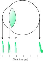


Fig. U1 Histogram of ultrasound reflections (or echoes) in the eye. Echoes from the various boundaries are given against total time, i.e. the time interval from the cornea to the boundary and back to the cornea. The velocity of the ultrasound waves in the eye is approximately 1550 m/s (it is 1641 m/s in the lens and 1532 m/s in the humours). In the above diagram the total time between the cornea and the retina is 32 μs. The length is then equal to 32/2 ✕ 10−6 ✕ 1550 ✕ 103 = 24
Millodot: Dictionary of Optometry and Visual Science, 7th edition. © 2009 Butterworth-Heinemann
ech·o
(ek'ō)1. Reverberating sound sometimes heard during chest auscultation.
2. In ultrasonography, acoustic signal received from scattering or reflecting structures or corresponding pattern of light on a cathose ray tube or ultrasonogram.
3. In magnetic resonance imaging, signal detected following an inverting pulse.
[G.]
Medical Dictionary for the Dental Professions © Farlex 2012
Patient discussion about echo
Q. My mother had a chest pain and she was sent for a TEE. When do you need a TEE and when a normal echo is fine? My mother had a chest pain few weeks ago. we were sure its a heart attack and went to the ER. There the doctors did some tests and she was sent for a (trans thoracic echocardiogram) TEE. I want to know when do you need a TEE and when you can do just a normal echocardiogram because the TEE was very painful for her and we want to know if ther was a better way.
A. The main difference between TEE and normal echo is that in TEE u put the transducer directly in the esophagus. The transducer is the same and the idea is to put it as close as possible to the heart.
As far as I know there are some heart situations the TEE is better for diagnosis that normal echo. Maybe your mom had one of those situations?
I can recommend you to ask the ER doctor. he will probably be able to give a better explanation for his choice
More discussions about echoAs far as I know there are some heart situations the TEE is better for diagnosis that normal echo. Maybe your mom had one of those situations?
I can recommend you to ask the ER doctor. he will probably be able to give a better explanation for his choice
This content is provided by iMedix and is subject to iMedix Terms. The Questions and Answers are not endorsed or recommended and are made available by patients, not doctors.
echoacousia
[ek″o-ah-koo´ze-ah]the subjective experience of hearing echoes after normally heard sounds.
Miller-Keane Encyclopedia and Dictionary of Medicine, Nursing, and Allied Health, Seventh Edition. © 2003 by Saunders, an imprint of Elsevier, Inc. All rights reserved.
ech·o·a·cou·si·a
(ek'ō-ă-kū'zē-ă),A subjective disturbance of hearing in which a sound appears to be repeated.
[echo + G. akouō, to hear]
Farlex Partner Medical Dictionary © Farlex 2012
ech·o·a·cou·si·a
(ek'ō-ă-kyū'zē-ă)A subjective disturbance of hearing in which a sound appears to be repeated.
[echo + G. akouō, to hear]
Medical Dictionary for the Health Professions and Nursing © Farlex 2012
ech·o·a·cou·si·a
(ek'ō-ă-kyū'zē-ă)Subjective hearing disturbance in which a sound repeats.
[echo + G. akouō, to hear]
Medical Dictionary for the Dental Professions © Farlex 2012
Latest Searches:
viscosity - viscosimetry - viscosimeter - viscose - viscometry - viscometer - Viscoheel - viscogel - viscoelasticity - viscoelastic - Viscoat - viscidity - viscid - visci - viscerum - viscerotropic - viscerotrophic - viscerotonia - viscerotomy - viscerotome -
- Service manuals - MBI Corp