cell
(sel) [L. cella, a chamber] 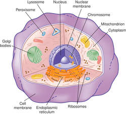

GENERALIZED HUMAN CELL AND ORGANELLES
The basic unit of life. A cell is a group of self-sustaining biochemical reactions that are isolated from the environment by a selectively permeable lipid membrane. Among the key reactions are those that maintain a stable intracellular concentration of ions; for mammalian cells, typical internal concentrations include 140 mM K+, 5-15 mM Na+, 5-15 mM Cl-, and a pH of 7.2, which can be significantly different from their concentrations outside the cell. Other key reactions move molecules and molecular complexes within the cell, sometimes changing the cell's shape. These reactions, along with many others, require energy, and the generation of energy by breaking apart preexisting hydrocarbon molecules ("food") is the job of glycolysis and other characteristic intracellular metabolic reactions. See: glycolysis; metabolism; mitochondrion.
Structure
Intracellular chemical reactions are controlled by enzymes that are organized in stable molecular complexes called organelles. The polymer-based organelles include centrioles and the cytoskeleton; nucleic acid-based organelles include ribosomes; and membrane-enclosed organelles include the nucleus, endoplasmic reticula, Golgi complexes, lysosomes, peroxisomes, mitochondria, and storage and transport vesicles. See: illustrationIndividual mammalian cells are usually microscopic, typically ranging from 5 µm to 50 µm in diameter. In humans, lymphocytes are small cells (~6 µm in diameter), columnar epithelial cells (10 µm x 20 µm) are medium-size cells, and mature ova (120-150 µm) are some of the largest cells.
Cell Division
In mammals, all new cells arise from existing cells through cell division, and an animal's growth results largely from increases in the number of its cells, most of which differentiate into specialized cell types to form the body's various tissues. Cell division involves two major processes: karyokinesis, the division of the nucleus, and cytokinesis, the division of the remainder of the cell. When generating somatic daughter cells, karyokinesis uses a process called mitosis, which produces daughter cells with a full complement of chromosomes. When generating germ cells, karyokinesis includes a process called meiosis, which produces daughter cells with half the normal number of chromosomes. See: meiosis and mitosis for illus.
A cell
Alpha cell of the pancreas.
accessory cell
A monocyte or macrophage that participates in the immune response.
See: antigen-presenting cell; macrophageacidophilic cell
Acidophil.
acinar cell
A cell present in the acinus of an acinous gland, e.g., of the pancreas.
adipose cell
Adipocyte..adult stem cell
A precursor cell that can also give rise to identical precursor cells: daughters of a stem cell can develop into a terminally-differentiated cell type or they can remain a stem cell. Adult stem cells are found in many tissues, such as bone marrow, brain, retina, skin, intestines, liver, testis, and pancreas.
Synonym: somatic stem cell See: embryonic stem celladventitial cell
A macrophage along a blood vessel, together with perivascular undifferentiated cells associated with it.
air cell
An air-filled sinus cavity in a bone.
alpha cell
1. An enteroendocrine cell that produces glucagon and is found in the pancreatic islets. Synonym: A cell
2. An acidophil of the adenohypophysis (anterior pituitary gland).
alveolar cell
1. In the lung, either of two types of epithelial cells lining the alveoli. Type I cells are simple thin squamous epithelial cells. Type II cells secrete pulmonary surfactant. Type II cells are smaller and more numerous than Type I cells.
2. In the mammary glands, the milk-secreting epithelial cells, which are activated during lactation.
amacrine cell
A modified nerve cell in the retina that has dendrites but no axon.
See: neuronameloblast cell
Ameloblast.
anterior horn cell
A somatic motor neuron that has its cell body in the ventral (anterior) horn of the gray matter of the spinal cord; its axon passes out through a ventral root and innervates skeletal muscle.
antigen-presenting cell
Abbreviation: APC
A cell that breaks down antigens and displays their fragments on surface receptors next to major histocompatibility complex molecules. This presentation is necessary for some T lymphocytes that are unable to recognize soluble antigens. Macrophages are the primary antigen-presenting cells, but B cells and dendritic cells also can act as APCs.
See: T cell; macrophage processing cellAPUD
The abbreviated name for an 'amine precursor uptake and decarboxylation cell'. These cells are the constituents of a diffuse neuroendocrine system and all have metabolic pathways that make and utilize serotonin (5-HT). APUD cells include chromaffin cells, enterochromaffin cells, and SIF cells as well as certain cells found in the parathyroid gland, thyroid gland, pituitary gland, hypothalamus, and placenta.
argentaffin cell
A cell in the epithelium of the stomach, intestines, and appendix that secretes serotonin.
astroglial cell
Astrocyte.atypical glandular cells
Abbreviation: AGC
An abnormal finding on a Pap test. This classification is divided into “favor neoplasia” or “not otherwise specified (NOS).” NOS is subdivided into endocervical or endometrial origin. Atypical endocervical cells are important because of their risk for significant disease.
Synonym: atypical glandular cells of undetermined significanceatypical glandular cells of undetermined significance
Abbreviation: AGUS
Atypical glandular cells.B cell
1. A lymphocyte that synthesizes and secretes antibodies. B lymphocytes originate and differentiate in the bone marrow and then populate the spleen, lymph nodes, and other lymphoid tissues. When exposed to an antigen, a B cell divides to form (a) plasma cells, which produce antigen-specific antibodies, and (b) a lesser number of memory B cells, which can quickly differentiate into plasma cells upon a second exposure to the original antigen. Antibody production is a key part of the humoral immune response of adaptive immunity. The humoral immune response is effective against bacteria, viruses, and other pathogens, and provides the rationale for vaccination. Synonym: B lymphocyte See: ; T cell;
2. Pancreatic beta cell.
band cell
The developing leukocyte at a stage at which the nucleus is not segmented.
basal cell
1. A rounded or cuboidal epithelial stem cell found in the bottom layer of pseudostratified epithelia, such as the epidermis and the lining of the airways of the lung.
2. Either of two types of cell found in the bottom layer of the olfactory epithelium; one type is a flattened "basal cell proper", and the other is a rounded stem cell called a globose cell.
3. A rounded stem cell found in the taste buds and a progenitor of the specialized taste receptor cells.
basket cell
1. Myoepithelial cell.
2. One of the nonspiny granule cells found in the cerebral cortex.
3. One of the small interneurons found in the outermost layer of the cerebellar cortex along with stellate cells.
basophilic cell
Basophil.
beta cell
1. Any of the insulin-secreting cells of the pancreas that constitute the bulk of the islets of Langerhans. Synonym: B cell
2. A basophil cell of the adenohypophysis (anterior pituitary gland).
Betz cell
See: Betz cellsbipolar cell
Bipolar neuron.
blast cell
1. A precursor cell for a specific cell type.
2. An immature cell of a specific type.
blood cell
Any cell normally found circulating in the blood stream. Blood cells include red cells and white cells; red cells generally remain inside blood vessels, while white cells can also more into the tissues outside the blood vessel walls.
See: blood for illus.bone cell
An osteoblast, osteoclast, or osteocyte.
bone marrow cell
Marrow cell.
brush cell
An epithelial cell found sparsely in the lining of the bronchial tree. The cell's surface has long stiff microvilli, and the cell has the appearance of an absorptive cell.
burr cell
An erythrocyte with 10 to 30 spicules distributed over the surface of the cell, as seen in heart disease, stomach cancer, kidney disease, and dehydration. Synonym: echinocyte
cancer cell
A cell present in a neoplasm and differentiated from normal tissue cells because of its degree of anaplasia, irregularity of shape, nuclear size, changes in the structure of the nucleus and cytoplasm, increased number of mitoses, and ability to metastasize.
capsule cell
Satellite cell.cartilage cell
Chondrocyte.castration cell
An enlarged and vacuolated basophil cell seen in the pituitary in gonadal insufficiency or following castration.
CD3 cell
T cell.
CD4 cell
Helper T cell.
CD8 cell
A suppressor T cell, e.g., a cytotoxic T cell.
CD 34+ cell
A cell with the CD34 protein on its surface membrane. Some CD34 cells that are hemopoietic stem cells can be separated out from peripheral blood.
cementoblast cell
Cementoblast.
cementocyte cell
Any of the cells trapped within cementum that maintain cementum as a living calcified tissue by their metabolic activity.
centroacinar cell
A duct cell of the pancreas more or less invaginated into the lumen of an acinus.
chalice cell
Goblet cell.chemoreceptor cell
Chemoreceptor.
chief cell
1. Any of the cells of the parathyroid gland that secretes the parathyroid hormone.
2. Any of the cells of the gastric glands that secretes pepsinogen.
chromaffin cell
A cell that produces, stores, and secretes catecholamines (dopamine and norepinephrine). Chromaffin cells are found in the medulla of the adrenal glands and in small clusters in the sympathetic ganglia.
chromophobe cell
Chromophobe.
Clara cell
A cuboidal epithelial cell found in the lining of the terminal and the respiratory bronchioles of the lungs. Clara cells are nonciliated, and they secrete surfactant, like the type II alveolar epithelial cells found deeper in the bronchial tree.
cleavage cell
Blastomere.clue cell
A vaginal epithelial cell, thickly coated with coccobacillary organisms. Clue cells are a hallmark of bacterial vaginosis.
columnar cell
An epithelial cell with height greater than its width.
columnar epithelial cell
Columnar cell.
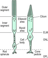
cone cell
A cell in the retina whose scleral end forms a cone that serves as a light receptor. Vision in bright light, color vision, and acute vision depend on the function of the cones. See: rod cell
contrasuppressor cell
A T cell that inhibits the activity of suppressor T cells. Although a contrasuppressor cell shares this functional capability with T helper cells, it is distinguished from other CD4+ cells by its other cell surface markers and the unique group of cytokines it produces.
cortical cell
A cell in the cortex of an organ, e.g., a neuron in the cerebral cortex.
corticotroph cell
Corticotroph.
cuboid cell
A cell – usually epithelial – with a height about equal to its width and depth.
cytotoxic cell
Cytotoxic T cell.
cytotoxic T cell
A CD8+ T lymphocyte that can destroy microorganisms directly through the release of perforin and proteolytic enzymes. These cells are particularly important in the defense against viruses, rejection of allografts, and, possibly, new malignant cells. Synonym: CD8 cell; cytotoxic cel; killer T cell
D cell
An enteroendocrine cell that produces somatostatin and is found in the pancreatic islets, stomach, and small intestine. Synonym: delta cell; somatostatin cell
daughter cell
A cell formed by cell division.
decoy cell
A cell found in the urine with inclusion bodies in its nucleus. It indicates infection with BK virus in renal transplant recipients.
delta cell
Pancreatic D cell.
dendritic cell
One type of antigen-presenting cell that helps T cells respond to foreign antigens. Dendritic cells are found in epithelial tissues and include the Langerhans' cells of the skin and the interdigitating cells in lymph nodes; they also circulate in the blood.
Downey cell
See: Downey celldust cell
A macrophage that migrates into the lumen of lung aveoli and ingests debris, particles of air pollution, and pathogens to keep the airspaces clear.
EC cell
1. An embryonal carcinoma cell, which is a cultured cell line.
2. An enterochromaffin cell that secretes substance P and is found in the stomach and small intestine.
effector cell
A cell that carries out the final response or function of a particular process. The main effector cells of the immune system, for example, are activated lymphocytes and phagocytes—the cells involved in destroying pathogens and removing them from the body.
See: leukocyteembryonic stem cell
Abbreviation: ES cell
A cell from the inner cell mass of the blastocyst (the 3-5 day old mammalian embryo) that can give rise to all the somatic cells of the body. Embryonic stem cells can be maintained as pure stem cell cultures.
See: adult stem cellendothelial cell
The type of epithelial cell that lines blood vessels and lymph vessels; these cells are usually squamous (flattened) and form sheets one layer thick. Endothelial cells are derived from mesenchyme cells of the embryo. A sheet of endothelial cells is called an endothelium.
See: endotheliumenterochromaffin cell
Abbreviation: EC cell
An enteroendocrine cell that produces serotonin and is found in the small intestine. Enterochromaffin cells are very similar to the cells, found throughout the peripheral sympathetic nervous system, that are called simply 'chromaffin cells'.
enteroendocrine cell
One of the scattered hormone-producing cells found in the pancreatic islets and throughout the gastrointestinal (mainly, small intestinal) mucosa.
ependymal cell
Any of the epithelial cells that form a one-cell-thick layer lining the ventricles and the central canal of the central nervous system. The ventricular (apical) surfaces of many ependymal cells are covered with cilia or microvilli. In most places, the ependymal layer does not have a basal lamina. Specialized regions of ependymal cells include the covering of the blood vessels and loose connective tissue of the choroid plexuses; here, the ependyma is specialized to secrete cerebrospinal fluid.
epithelial cell
Any of the cells forming the cellular sheets that cover surfaces, both inside and outside the body. Epithelial cells are closely packed and take on polyhedral shapes, from tall (columnar) through squat (cuboidal) to flat (squamous). Epithelial cells adhere strongly to one another, and one of their surfaces -- the basal surface -- sticks firmly to a thin extracellular film of fibrils called a basal lamina. A sheet of epithelial cells derived from embryonic epithelia (the ectoderm or the endoderm) is called an epithelium. See: epithelium
ethmoidcell
Ethmoid sinus.
ethmoid air cell
Ethmoid sinus.
eukaryotic cell
The type of cell composing multicellular, as well as a number of unicellular, organisms. Unlike prokaryotic cells, eukaryotic cells have many of their intracellular functions organized within structures called organelles. Some organelles -- notably, the nucleus, which contains the DNA -- are enclosed by intracellular membranes.
F cell
An enteroendocrine cell that produces pancreatic polypeptide and is found in the pancreatic islets.
fat cell
Adipocyte.
flame cell
A bone marrow cell with a bright red cytoplasm, occasionally found in the marrow of patients with multiple myeloma.
flow cell
An optical cell used in photometers and cell counters, through which the sample and any standards are passed for detection and measured or counted by optical or electrometric means.
See: cytometryfoam cell
A cell that contains vacuoles; a lipid-filled macrophage.
follicle cell
Follicular cell.
follicular cell
1. The secretory cell of the thyroid gland; it produces the thyroid hormones, T3 (triiodothyronine) and T4 (tetraiodothyronine or thyroxine).
2. Any of the flattened somatic cells that form a monolayer around each primary oocyte in the ovary. After puberty, when an oocyte matures, during a monthly cycle, its follicular cells divide, become cuboidal, and form a multilayered coating for the oocyte; at this stage, the follicular cells are called granulosa cells.
folliculostellate cell
A supporting cell in the adenohypophysis (anterior pituitary gland); it produces bioactive peptides, including growth factors and cytokines.
foreign body giant cell
Giant cell.
G cell
An enteroendocrine cell found in the stomach that produces the hormone gastrin.
ganglion cell
1. Any neuron whose cell body is located within a ganglion.
2. A neuron of the retina of the eye whose cell body lies in the ganglion cell layer. The axons of ganglion cells form the optic nerve.
germ cell
A cell whose function is to reproduce the organism. Early in development, primordial germ cells are found in the genital ridges of the embryo. Later, in the testis, the primordial germ cells are called spermatogonia, and in the ovary, they are called oogonia. When they mature, the germ cells (i.e., spermatogonia and oogonia) differentiate into haploid gametes (i.e., spermatozoa and ova).
Synonym: primordial germ cellgiant cell
1. A multinucleated phagocyte created by several individual macrophages that have merged around a large pathogen or a substance resistant to destruction, such as a splinter or surgical suture. See: granuloma; tuberculosis
2. A large multinucleated (40-60 nuclei) tumor cell characteristic of certain bone and tendon tumors.
3. A large multinucleated cell that invades the walls of the aorta and its major branches in giant cell arteritis.
glial cell
One of three types of nonneuronal cell in the central nervous system: astrocytes, oligodendrocytes, and microglial cells. SYN neuroglial cell
Synonym: neuroglial cellgitter cell
A macrophage present at sites of brain injury. The cells are packed with lipoid granules from phagocytosis of damaged brain cells.
See: microgliaglobus cell
One of the two varieties of basal cell found in the olfactory epithelium. It is a rounded neuroblast or neural stem cell for the olfactory receptor cells.
goblet cell
A mucous cell sitting between nonsecretory cells, such as is found in the intestinal epithelium.
Golgi cell
See: Golgi, Camillogonadotroph cell
Gonadotroph.
graft facilitating cell
Any of a group of CD8 positive, t-cell receptor negative cells that help donated bone marrow engraft in the recipient.
granule cell
1. Any of the small neurons that pack the granular cell layer of the cerebellar cortex, immediately below the Purkinje cell layer. Granule cells receive inputs (mossy fibers) from the spinal cord and brainstem (except the inferior olive). Axons of granule cells run perpendicular to the Purkinje cell dendrites, on which they synapse.
2. Any of the neurons of the cerebral cortex that are not pyramidal cells. Cortical granule cells are categorized as spiny or nonspiny. Synonym: stellate cell
3. A small axon-less neuron found in the olfactory bulb.
granulosa cell
One of the many cuboidal cells that surround and nurture the maturing oocyte.
See: follicular cell (2)gustatory cell
Taste cell.
hair cell
An epithelial cell possessing stereocilia in the maculae, cristae ampullaris, and the organ of Corti. These cells are receptors for the senses of position and hearing.
heart failure cell
A red-colored (from ingested red cells) lung macrophage often found in the sputum of patients with congestive heart failure.
HeLa cell
A line of human epithelial cells that grows well in culture. It is an immortal cancer cell that has been maintained in continuous tissue cultures for decades from a patient with carcinoma of the cervix. It is named for the first two letters of the patient's first and last names, Henrietta Lacks. HeLa cells have been used in thousands of experiments on cell growth, differentiation, and cancer, and in virology, pharmacology, and other fields.
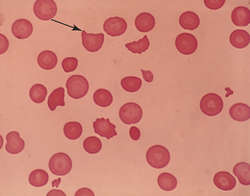
HELMET CELLS: (Orig. mag. ×640)
helmet cell
A schistocyte or fragmented blood cell, seen in hemolytic anemias.
See: illustrationhelper cell
A type of T lymphocyte whose surface is marked by CD4 receptors; it is involved in both cell-mediated and antibody-mediated immune responses. It secretes cytokines that stimulate the activity of B cells and other T cells and binds with class II histocompatibility antigens, which are processed by macrophages and other antigen-presenting cells. Synonym: helper T cell See: antigen processing; T cell; cell-mediated immunity
helper T cell
Helper cell.
hematopoietic stem cell
A progenitor cell in the bone marrow that can replicate itself as well as produce precursor cells of the various blood cell lineages.
hilus cell
An androgen-producing cell found in the ovarian hilum. It is analogous to the male Leydig cell.
holly leaf cell
A cell found in blood smears of persons with sickle cell anemia.
horizontal cell
A neuron of the inner nuclear layer of the retina. The axons of these cells run horizontally and connect various parts of the retina.
Hürthle cell
See: Hürthle cellhybridoma cell
See: hybridomahyperchromatic cell
A cell that contains more than the normal number of chromosomes and hence stains more densely.
I cell
An enteroendocrine cell that produces the enzyme cholecystokinin-pancreozymin (pancreaticozymin) and is found in the small intestine.
interdigitating cell
A type of antigen-presenting cell found in lymph nodes and lymphoid tissue.
interstitial cell
Any of the many cells found in connective tissue of the ovary, in the seminiferous tubules of the testes, and in the medulla and cortex of the kidney. The cells in the testes and ovaries produce hormones such as testosterone and estrogen.
intestinal absorptive cell
In the small intestine, any of the tall columnar cells topped with a brush border made of thousands of microvilli.
islet cell
A cell of the islets of Langerhans of the pancreas.
juvenile cell
The early developmental form of a leukocyte.
juxtaglomerular cell
A modified smooth muscle cell in the wall of the afferent arteriole leading to a glomerulus of the kidney. This type of cell secretes renin when blood pressure decreases to activate the renin-angiotensin mechanism, which increases sodium retention, thus elevating the blood pressure.
K cell
An enteroendocrine cell that produces gastric inhibitory peptide (glucose-dependent insulinotropic peptide) and is found in the small intestine. This peptide stimulates the beta cells of the pancreas to secrete insulin.
killer cell
Natural killer cell.
killer T cell
Cytotoxic T cell.Kulchitsky cell
An APUD cell found in the lung.
Kupffer cell
See: Kupffer cellL cell
An enteroendocrine cell that produces glucagon-like peptide-1 and is found in the small intestine. This peptide signals the pancreas to secrete insulin after a meal.
labile cell
A cell that is always mitotically active, such as the epithelial cells lining the stomach and the stem cells in the red bone marrow.
lactotroph cell
Lactotroph.
LAK cell
Abbreviation for lymphokine-activated killer cell. These natural killer cells, obtained from the patient's blood, have been activated in culture with interleukin-2.LAK cells; the cells can then be used to treat patients with solid malignant tumors.
Langerhanscell
A type of dendritic antigen-presenting cell that typically resides in the skin.
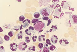
L.E. CELL (center): (Orig. mag. ×1000)
L.E. cell
Historically, an abbreviation for
lupus erythematosus cell, a polymorphonuclear leukocyte that contains the phagocytized nucleus of another cell. It is characteristic but not diagnostic of lupus erythematosus.
This distinctive cell may form when the blood of patients with systemic lupus erythematosus is incubated and further processed according to a specified protocol. The plasma of some patients contains an antibody to the nucleoprotein of leukocytes. These altered nuclei, which are swollen, pink, and homogeneous, are ingested by phagocytes. These are the L.E. cells. The ingested material, when stained properly, is lavender and displaces the nucleus of the phagocyte to the inner surface of the cell membrane. The L.E. cell phenomenon can be demonstrated in most patients with systemic lupus erythematosus but is not essential for diagnosis.
Leydig cell
See: Leydig's celllittoral cell
A macrophage found in the sinuses of lymphatic tissue.
liver cell
Hepatocyte.
lutein cell
A cell of the corpus luteum of the ovary that contains fatty yellowish granules. Granulose lutein cells are hypertrophied follicle cells; these lutein (paralutein) cells develop from the theca interna.
lymphoid cell
An obsolete term for lymphocyte.
lymphokine-activated killer cell
LAK cell.
M cell
1. A microfold cell, which is a cell in the gastrointestinal epithelium covering patches of lymphoid tissue. M cells transport antigens from the intestinal lumen to the underlying lymphoid tissues for recognition and processing.
2. An APUD cell that produces melanotropin and is found in the pituitary gland.
macroglial cell
An astrocyte or an oligodendrocyte.
marrow cell
Bone marrow cell.
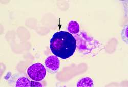
MAST CELL
mast cell
A large tissue cell resembling a basophil, which is essential for inflammatory reactions mediated by immunoglobulin E (IgE) but does not circulate in the blood. Mast cells are present throughout the body in connective tissue, but are concentrated beneath the skin and the mucous membranes of the respiratory and digestive tracts. Mast cells are covered with IgE molecules, which bind with foreign antigens and stimulate degranulation, releasing such mediators as histamine, prostaglandins, leukotrienes, and proteinases from densely packed granules within the cytoplasm. These mediators produce type I (immediate) hypersensitivity reactions (e.g., urticaria, allergic rhinitis, asthma, angioedema, and systemic anaphylaxis). See: illustration
mastoid cell
Mastoid air cell.
mastoid air cell
Any of the variable-sized, air-filled sinuses inside the mastoid antrum. About 20% of adult skulls have no mastoid air cells.
matrix stem cell
A stem cell derived from Wharton's jelly.
memory cell
A cell derived from B or T lymphocytes that can quickly recognize a foreign antigen to which the body has been previously exposed. Memory T cells stimulate T helper lymphocytes and cytotoxic T cells; memory B cells stimulate the production of antigen-specific antibodies by B plasma cells. Both types of memory cells survive for years, providing a durable adaptive immune response against foreign antigens.
mesenchyme cell
One of the two basic somatic cell lineages -- the other being epithelial cells. In contrast to epithelial cells, mesenchyme cells are not polarized and are frequently motile. In the early embryo, mesenchyme cells fill many of the spaces enclosed by epithelia. Later, mesenchyme cells will secrete the space-filling extracellular matrix molecules, such as collagen and glycoproteins, that characterize connective tissue.
mesenchymal stem cell
A stem cell found in connective tissue and capable of producing cells of the connective tissue lineages, such as cartilage, bone, muscle, and fat cells.
mesothelial cell
The type of epithelial cell that lines serous (pleural, peritoneal, and pericardial) cavities, blood vessels, and lymph vessels; these cells are usually squamous (flattened) and form sheets one layer thick. Mesothelial cells are derived from mesenchyme cells of the embryo. A sheet of mesothelial cells is called a mesothelium.
See: mesotheliummicroglial cell
A small glial cell of the central nervous system and retina. Microglia have spiky branched processes and are arranged homogeneously throughout the brain and spinal cord. They are activated by disease and injury, after which they become phagocytic and sometimes resume their embryonic motility like a macrophage.
mitral cell
One of the two principal neurons of the olfactory bulb -- the other being the tufted cell. In a complex synaptic formation called a glomerulus, each of the mitral and tufted cells receives synaptic inputs from axons of the olfactory nerve. The axons of the mitral and tufted cells form the olfactory tract and synapse in the olfactory cortex.
mossy cell
An astrocyte or other glial cell with many branching processes. See: neuroglia
mother cell
A cell that gives rise to similar cells through fission or budding. Synonym: parent cell
mucosal cell
Any cell in a mucosal epithelium.
mucous cell
An epithelial cell that secretes mucus and IgA antibodies. Mucous cells and serous cells are the two varieties of secretory cells found in exocrine glands.
Synonym: mucus cellmucus cell
Mucous cell.
multinucleated giant cell
Giant cell.
multipolar cell
Multipolar neuron.
muscle cell
See: musclemyeloid cell
Any white blood cell other than lymphocytes.
myeloma cell
A cell present in the bone marrow of patients with multiple myeloma.
myoepithelial cell
A smooth muscle cell found in some epithelia; it lies between glandular cells and the basal lamina of sweat, mammary, lacrimal, and salivary glands.
Synonym: basket cellnatural killer cell
Abbreviation: NK cell
A large granular lymphocyte – a defensive cell of innate immunity – that bonds to cells and lyses them by releasing cytotoxins. Natural killer cells are null cells, lymphocytes that do not have B cell or T cell surface markers, and they can be activated without previous antigen exposure. NK cells destroy cells infected with viruses and some types of tumor cells in cultures. They also secrete gamma interferon (INF?), tumor necrosis factor alpha (TNFa), and granulocyte-macrophage colony-stimulating factor (GMCSF), enhancing the effect of T lymphocytes.
Synonym: killer cellnerve cell
Neuron.neural crest cell
Embryonic cells of the neuron-glia lineage that form along the ridges (neural folds) of the neural plate and that migrate into the developing organism to produce a variety of tissues. The migratory ability of these embryonic epithelial cells is similar to the motility of mesenchyme cells; this has led neural crest cells to be called mesectodermal cells. In the neural lineage, neural crest cells give rise to the dorsal root ganglia, the placodes that will develop into the olfactory and auditory sensory organs, the pituitary gland, the peripheral autonomic nervous system, and the neurenteric and APUD cells. In the glial lineage, neural crest cells give rise to Schwann cells and other peripheral satellite cells. In addition, neural crest cells of the cranial region give rise to certain facial connective tissue, including the bones of the nasal cavities, the roof of the mouth, and the sella turcica.
neuroglial cell
Glial cell.
Niemann-Pick cell
See: Niemann-Pick cellNK cell
Natural killer cell.
nonstem cell
Any cell found in the bone marrow that cannot reconstitute the marrow or give rise to more differentiated blood cells.
null cell
A large lymphocyte without the cell markers of either a T cell or a B cell. Natural killer cells are examples of null cells.
odontoblast cell
Odontoblast.
oligodendroglial cell
Oligodendrocyte.
olfactory cell
Olfactory receptor cell.
olfactory receptor cell
A cell of the olfactory mucosa that has receptors for the sense of smell. Olfactory cells are continuously replaced from stem cells throughout adult life.
Synonym: olfactory cellosteoprogenitor cell
Any of the mesenchyme precursor cells committed to the bone lineage and capable of producing osteoblasts and osteocytes. Osteoprogenitor cells are found in bone, bone marrow, and other connective tissue.
oxyntic cell
A parietal cell of the gastric glands; it produces hydrochloric acid and the intrinsic factor.
parabasal cell
An abnormal but not malignant cell seen in some cytologic specimens obtained during Papanicolaou tests (Pap tests). It is found in women with vaginal atrophy, in some postpartum women, some women suffering from anorexia or starvation, and some who have used progesterone for contraception.
parent cell
Mother cell.phalangeal cell
One of the cells supporting the hair cells of the organ of Corti. These cells form several rows of outer phalangeal cells (Deiters' cells) and a single row of inner phalangeal cells.
phantom cell
See: red cell ghostpigment cell
Any cell that normally contains pigment granules.
plasma cell
A cell derived from a B lymphocyte that has been sensitized to a specific foreign antigen and produces antibodies to that particular antigen. It may be found in the blood or in tissue fluid. Synonym: plasmacyte
postganglionic cell
Postganglionic neuron.
PP cell
An enteroendocrine endocrine cell found in the pancreatic islets that produces pancreatic polypeptide.
pre-B cell
The immediate precursor of a lymphocytic B cell.
preganglionic cell
Preganglionic neuron.
prickle cell
A cell with spiny processes that connect with similar processes of adjoining cells. These are found in the stratum spinosum of the keratinized epithelium of the epidermis.
primary cell
In physical therapy, a device consisting of a container, two solid conducting elements, and an electrolyte for the production of electric current by chemical energy.
primordial cell
Primordial germ cell.
primordial germ cell
A germ cell before it begins its maturation into a haploid gamete.
Synonym: primordial cellprogenitor cell
A cell (sometimes a stem cell) that produces cells of a particular lineage, e.g., a neuroblast.prokaryotic cell
The form of cell composing many primitive unicellular organisms, such as bacteria. Prokaryotic cells do not have nuclei, which are partitioned by an intracellular membrane; instead the DNA forms one main coil in the cell cytoplasm.
Purkinje cell
See: Purkinje, Johannes E. vonpus cell
A leukocyte present in pus. Pus cells are often degenerated or necrotic.
pyramidal cell
A large, common neuron found in the cerebral cortex. Pyramidal cells are flask-shaped or triangular, and, in the parts of the cortex with six layers, they occupy the fifth layer. Pyramidal cell dendrites project up into the most superficial layer of the cortex, while pyramidal cell axons run in the opposite direction, i.e., downward and out of the cortex.
Synonym: pyramidal neuronradial glial cell
A structural macroglial cell that is a key component of the developing nervous system. Radial glial cells first appear in the neural tube, where their cell bodies are suspended between two thin cell processes; the apical process attaches to the inner (ventricular) surface of the neural tube, and the basal process attaches to the outer (pial) surface. Early in development, neuroblasts migrate radially along the scaffolding formed by the radial glial cell processes, and growing axons may follow the scaffolding longitudinally. Later, many radial glial cells retract their processes and differentiate into astrocytes.
red cell
A small cell that is filled with hemoglobin, has no nucleus, and is shaped like a biconcave disc. Red cells transport oxygen to tissues and carbon dioxide to the lungs. Individual red cells have a life span of 3-4 months, and new red cells are continually being produced in the bone marrow. In a healthy person, 99% of the cells circulating in the blood are red cells.
Synonym: erythrocyte; red blood cell; red blood corpuscle See: erythrocytered blood cell
Abbreviation: RBC
Red cell.
Renshaw cell
See: Renshaw cellresting cell
1. A cell that is not dividing. See: interphase
2. A cell not performing its normal function (i.e., a nerve cell that is not conducting an impulse or a muscle cell that is not contracting).
reticular cell
1. An undifferentiated cell of the spleen, bone marrow, or lymphatic tissue that can develop into one of several types of connective tissue cells or into a macrophage.
2. A cell of reticular connective tissue. See: reticular tissue
reticuloendothelial cell
An out-of-date term for a cell of the mononuclear phagocytic system.
Rieder cell
See: Rieder cellrod cell
A cell in the retina of the eye whose scleral end is long and narrow, forming a rod-shaped sensory receptor. Rods are stimulated by light and are essential for vision in dim light. See: cone cell
rosette cell
A rose-shaped cluster of phagocytes surrounding lysed nuclear material or red blood cells. Rosette cells occur frequently in blood in which L.E. cells are present. Rosette cells are not diagnostic of lupus erythematosus. See: L.E. cell
Rouget cell
See: Rouget cellS cell
An enteroendocrine cell that produces secretin and is found in the small intestine.
satellite cell
1. A stem cell associated with skeletal muscle that may form a limited number of new muscle cells after injury.
2. One of the neuroglia cells enclosing the cell bodies of sensory neurons in spinal ganglia. Synonym: capsule cell
scavenger cell
A phagocyte that cleans up disintegrating tissues or cells.
Schwann cell
See: Schwann cellsegmented cell
A segmented neutrophil (i.e., one with a nucleus of two or more lobes connected by slender filaments).
selenoid cell
See: red cell ghostsensory cell
A cell that when stimulated gives rise to nerve impulses that are conveyed to the central nervous system.
septal cell
A type II alveolar cell that secretes pulmonary surfactant; it is adjacent to a septum of the alveoli.
serous cell
An epithelial cell that secretes a watery fluid containing proteins, glycoproteins, and often antibodies (IgA, IgG, and IgM). Serous cells and mucous cells are the two varieties of secretory cells found in exocrine glands.
Sertoli cell
See: Sertoli cellsex cell
Gamete.
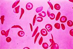
SICKLED CELLS IN SICKLE CELL DISEASE
sickle cell
An abnormal erythrocyte shaped like a sickle. Synonym: drepanocyte See: anemia, sickle cell
illustrationsignet-ring cell
A vacuolated cell with the nucleus off center. Mucus-secreting adenocarcinomas usually contain these cells.
skeletal muscle cell
See: musclesmooth muscle cell
See: musclesomatic cell
Any cell that is not a germ cell.
somatic stem cell
Adult stem cell.
somatostatin cell
D cell.
spiculed red cell
Crenated red blood cells with surface projections. In most instances, this is a normal variation in red cell equilibrium and is reversible.
See: acanthocytespider cell
Astrocyte.squamous cell
A flat epithelial cell.
Synonym: squamous epithelial cellsquamous epithelial cell
Squamous cell.
stellate cell
1. Granule cell of the cerebral cortex.
2. One of the small interneurons found in the outer layer of the cerebellar cortex along with basket cells.
stellate reticuloendothelial cell
A Kupffer cell, one of the macrophages that line the sinusoids of the liver.
stem cell
An embryonic stem cell or an adult stem cell.
Sternberg-Reed cell
See: Reed-Sternberg cellstipple cell
A red blood cell that contains small basophilic-staining dots. It is seen in lead poisoning, malaria, severe anemia, and leukemia.
striated muscle
See: musclesuppressor T cell
A subpopulation of regulatory T lymphocytes that develop in the thymus gland, that slows or stops a specific immune response.
sustentacular cell
A supporting cell, as in the acoustic macula, organ of Corti, olfactory epithelium, taste buds, or testes.
sympathicotrophic cell
One of the large epithelial cells that occur in groups in the hilus of the ovary. They are thought to be chromaffin cells.
sympathochromaffin cell
A chromaffin cell of ectodermal origin present in the fetal adrenal gland. Sympathetic and medullary cells originate from these cells.
syncytial giant cell
Giant cell.
T cell
A lymphocyte that responds to specific antigens, with the assistance of antigen-presenting cells (APCs). T cells arise in the bone marrow and migrate to the thymus gland, where they mature; then they circulate between blood and lymph, serving as one of the primary cells of the adaptive immune response. Immature T cells are called thymocytes. Mature T cells are antigen specific. Their surface receptors (T cell receptors, abbrev. TCRs) respond only to a single antigen. T cells are further categorized using another family of surface protein markers called clusters of differentiation (CDs). All T cells have a CD3 marker. Additional markers differentiate the subclasses of T cells. CD4 T helper cells serve primarily as regulators, secreting cytokines that stimulate the activities of other white blood cells. CD8 T cells (cytotoxic T cells) directly lyse (kill) organisms, an important defense against viruses; most CD8 T cells also produce gamma interferon (INF?), one of the strongest stimulators of macrophage activity. Synonym: T lymphocyte See: immune response; lymphocyte; immunological surveillance; T-cell receptor
A T cell can only recognize the "foreignness" of antigens after they have been modified by macrophages and other antigen-presenting cells (APCs). After this, T cells dominate the adaptive immune response by mobilizing B cells and other T cells of the cell-mediated immune pathways. T cells are responsible for type IV hypersensitivity reactions, such as graft rejection, and for tumor cell recognition and destruction.
See: cytokine; cell-mediated immunity
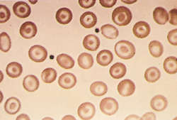
TARGET CELLS: In hemoglobin C disease (×600)
target cell
1. . An erythrocyte with a dark rounded central area surrounded by a lightly stained clear ring, which in turn is surrounded by a dense ring of peripheral cytoplasm. It is present in certain blood disorders, such as thalassemia, and in patients who have no spleen. See: hemoglobin C disease for illus Synonym: codocyte; leptocyte
2. . The cell at which a signal (e.g., hormone or nerve impulse) is aimed.
illustrationtart cell
A phagocyte that has ingested the unaltered nuclei of cells. These nuclei can be observed unchanged within the phagocytes.
taste cell
Any of the neuroepithelial cells within a taste bud that are receptors for the sense of taste. Each possesses on the free surface a short gustatory hair that projects through the inner taste pore.
Synonym: taste receptor celltaste receptor cell
taste cell.tear-drop cell
An abnormally shaped blood cell, sometimes found on blood smears of patients with bone marrow fibrosis, iron deficiency, or thalassemias.
Synonym: dacrocytetendon cell
Any of the fibroblasts of white fibrous connective tissue of tendons arranged in parallel rows.
terminally differentiated cell
A cell sufficiently committed to a particular function that it can no longer divide, e.g. a red cell.
thymic epithelial cell
The epithelial cells that form the internal scaffolding of the thymus. These cells vary in shape and size but generally align in sheets and cords, partitioning the thymus into islands of close-packed lymphocytes in the organ's cortex. Thymic epithelial cells are not simply structural and they interact actively with adjacent lymphocytes.
thymus cell
Any cell characteristic of the thymus, including thymic epithelial cells and thymocytes (thymic lymphocytes).
thyroid cell
Any cell characteristic of the thyroid gland, but usually referring to a thyroid follicular cell.
totipotent cell
An undifferentiated embryonic cell that has the potential to develop into any type of cell.
Touton giant cell
See: Touton celltransitional cell
The stretchable epithelial cells that compose the transitional epithelium (uroepithelium), which lines most of the urinary tract. Transitional cells are strongly interconnected. They are cuboidal when not under pressure, and they become flattened and squamous when stretched. Transitional epithelia are 4-6 cells thick, and the top transitional cells -- those on the lumenal surface -- fuse to become larger and polyploid.
trophoblast cell
One of the epithelial cells forming the surface of the spherical blastocyst stage embryo. Trophoblast cells are destined to give rise to many of the extraembryonic tissues.
tufted
See: mitral cellTürk irritation cell
See: Türk irritation cellTzanck cell
See: Tzanck cellundifferentiated cell
A cell resembling an embryonic cell in that it does not have the specific morphologic or functional characteristics of any particular adult cell type.
unipolar cell
A cell with a single cell process.
Vero cell
A lineage of cells used in cell cultures and isolated from kidney epithelial cells of the African green monkey (Cercopithecus aethiops).
visual cell
A rod cell or cone cell of the retina.
wandering cell
A rarely used term for a cell (such as a macrophage) that moves like an ameba.
white cell
Leukocyte.white blood cell
Abbreviation: WBC
Leukocyte.illustrationMedical Dictionary, © 2009 Farlex and Partners