Medical term:
corneae
cornea
[kor´ne-ah]the clear, transparent anterior covering of the eye (see also color plates). The cornea is subject to injury by foreign bodies in the eye, bacterial infection, and viral infection, especially by the herpesvirus that causes herpes simplex. The herpesvirus that causes herpes zoster (shingles) can also infect the cornea. Prompt treatment of any corneal injury or infection is essential to avoid ulceration and loss of vision.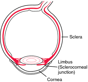

Cornea. From Dorland's, 2000.
Miller-Keane Encyclopedia and Dictionary of Medicine, Nursing, and Allied Health, Seventh Edition. © 2003 by Saunders, an imprint of Elsevier, Inc. All rights reserved.
cor·ne·a
(kōr'nē-ă), [TA]The transparent tissue constituting the anterior sixth of the outer wall of the eye, with a 7.7-mm radius of curvature as contrasted with the 13.5 mm of the sclera; it consists of stratified squamous epithelium continuous with that of the conjunctiva, a substantia propria, substantially regularly arranged collagen imbedded in mucopolysaccharide, and an inner layer of endothelium. It is the chief refractory structure of the eye.
[L. fem. of corneus, horny]
Farlex Partner Medical Dictionary © Farlex 2012
cornea
(kôr′nē-ə)n.
The transparent convex anterior portion of the outer fibrous coat of the eyeball that covers the iris and the pupil and is continuous with the sclera.
cor′ne·al (-əl) adj.
The American Heritage® Medical Dictionary Copyright © 2007, 2004 by Houghton Mifflin Company. Published by Houghton Mifflin Company. All rights reserved.
cor·ne·a
(kōr'nē-ă) [TA]The transparent tissue constituting the anterior sixth of the outer wall of the eye, with a 7.7-mm radius of curvature as contrasted with the 13.5-mm radius of the sclera. It consists of stratified squamous epithelium continuous with that of the conjunctiva, a substantia propria, regularly arranged collagen imbedded in mucopolysaccharide, and an inner layer of endothelium. It is the chief refractory structure of the eye.
[L. fem. of corneus, horny]
Medical Dictionary for the Health Professions and Nursing © Farlex 2012
cornea
The outer, and principle, lens of the eye through which the coloured iris with its central hole (the pupil) can be seen. The cornea performs most of the focusing of the eye. Fine adjustment (ACCOMMODATION) is done by the internal crystalline lens.Collins Dictionary of Medicine © Robert M. Youngson 2004, 2005
cornea
that part of the SCLERA at the front of the eye of vertebrates overlaying the iris and lens. It is a transparent layer of EPITHELIUM and CONNECTIVE TISSUE through which light enters the eye and is refracted so that the lens can then focus it on the retina. The cornea contains no blood vessels, oxygen coming directly from air dissolved in tear secretions, and is therefore easy to transplant as no tissue-typing is required.Collins Dictionary of Biology, 3rd ed. © W. G. Hale, V. A. Saunders, J. P. Margham 2005
Cornea
Clear, bowl-shaped structure at the front of the eye. It is located in front of the colored part of the eye (iris). The cornea lets light into the eye and partially focuses it.
Mentioned in: Cataract Surgery, Corneal Abrasion, Corneal Transplantation, Exophthalmos, Eye Cancer, Eye Examination, Eye Glasses and Contact Lenses, Foreign Objects, Glaucoma, Hyperopia, Inclusion Conjunctivitis, Laser Surgery, Myopia, Photorefractive Keratectomy and Laser-Assisted In-Situ Keratomileusis, Pinguecula and Pterygium, Radial Keratotomy, Sjögren's Syndrome, Starvation, Tarsorrhaphy, Tissue Typing, Trabeculectomy, Trachoma, Uveitis, Visual Impairment, Vitamin A Deficiency
Gale Encyclopedia of Medicine. Copyright 2008 The Gale Group, Inc. All rights reserved.
cornea
The transparent anterior portion of the fibrous coat of the globe of the eye. It has a curvature somewhat greater than the rest of the globe, so a slight furrow marks its junction with the sclera. Looked at from the front the cornea is about 12 mm horizontally and 11 mm vertically. It is the first and most important refracting surface of the eye, having a power of about 42 D. The anterior surface has a radius of curvature of about 7.8 m, the posterior surface 6.5 mm, and the central thickness is about 0.5 mm. It consists of five layers, starting from the outside: (1) the stratified squamous epithelium; (2) Bowman's layer; (3) the stroma (substantia propria); (4) Descemet's membrane; and (5) the endothelium. The cornea is avascular, receiving its nourishment by permeation through spaces between the lamellae. The sources of nourishment are the aqueous humour, the tears and the limbal capillaries. The cornea is innervated by the long ciliary and other nerves of the surrounding conjunctiva, which are all branches of the ophthalmic division of the trigeminal nerve. Innervation is entirely sensory. Within the cornea there are only unmyelinated nerve endings. The density of nerves in the cornea is very high, making it the most sensitive structure in the body. The major structural component of the cornea is collagen, mostly type I, and most of the ground substance between the collagen fibrils in the corneal stroma is proteoglycans whose core proteins bind with keratan sulfate and dermatan sulfate (chondroitin sulfate B). The cornea owes its transparency to the regular arrangement of the collagen fibrils, but any factor that affects this lattice structure (e.g. swelling, pressure) results in a loss of transparency. The cornea contains some 78% water, some 15% collagen and some 5% of other proteins (Fig. C18). See endothelial bedewing; corneal stroma; corneal topography; dellen; deturgescence; dyskeratosis; glycosaminoglycan; keratitis; keratomycosis; Hudson-Stahli line; Bowman's layer; Descemet's membrane; microcornea; specular microscope; optical zone of cornea; pachometer; Maurice's theory; Hurler's syndrome; videokeratoscope.
conical cornea See keratoconus.
cornea farinata A bilateral corneal degeneration characterized by faint dust-like opacities in the deep stroma. They do not impair vision and are usually age-related.
cornea guttata Dystrophy of the endothelial cells of the cornea which may result from corneal trauma, cataract surgery, keratic precipitates, tonography, ageing, continuous contact lens wear, or as part of the early stages of Fuch's endothelial dystrophy (a disease associated with ageing and with females more than males). It is seen clinically by slit-lamp examination as black spherules in the endothelial pattern. The condition is bilateral, although one eye may be affected more than the other. As the condition progresses the cornea becomes oedematous with a consequent loss of vision and eventually turns into bullous keratopathy. If the degenerated cells are located at the periphery of the cornea they are called Hassall-Henle bodies and are of no clinical significance except as an indication of ageing. Syn. corneal guttae; endothelial corneal dystrophy. See specular reflection illumination; keratic precipitates.
optical zone of cornea See optical zone of cornea.
cornea plana A rare, congenital, usually bilateral condition in which the corneal curvature is flatter than normal with a significant decrease in refractive power. The eye is usually hyperopic with a shallow anterior chamber often resulting in angle-closure glaucoma. There is some degree of peripheral scleralization and it is closely associated with sclerocornea.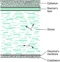

Fig. C18 Diagram showing the various layers of the cornea (k, keratocytes)
Millodot: Dictionary of Optometry and Visual Science, 7th edition. © 2009 Butterworth-Heinemann
cor·ne·a
(kōr'nē-ă) [TA]Transparent tissue constituting the anterior sixth of the outer wall of the eye, with a 7.7-mm radius of curvature.
[L. fem. of corneus, horny]
Medical Dictionary for the Dental Professions © Farlex 2012
Patient discussion about Cornea
Q. Has anyone had experience with a corneal transplant because of keratoconus?
A. my uncle had to do a transplant- it took 5 weeks until he could see anything , another year to get his vision straightened up. but now he is fine! i know that he looked for information in the "National Keratoconus Foundation". they were very helpful (and nice!), they have a website with information on all forms of treatment:
http://www.nkcf.org/
good luck :)
More discussions about Corneahttp://www.nkcf.org/
good luck :)
This content is provided by iMedix and is subject to iMedix Terms. The Questions and Answers are not endorsed or recommended and are made available by patients, not doctors.
Latest Searches:
viscosity - viscosimetry - viscosimeter - viscose - viscometry - viscometer - Viscoheel - viscogel - viscoelasticity - viscoelastic - Viscoat - viscidity - viscid - visci - viscerum - viscerotropic - viscerotrophic - viscerotonia - viscerotomy - viscerotome -
- Service manuals - MBI Corp