Medical term:
csf
cerebrospinal
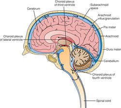
[ser″ĕ-bro-spi´nal]The cerebrospinal fluid aids in the protection of the brain, spinal cord, and meninges by acting as a watery cushion surrounding them to absorb the shocks to which they are exposed. There is a blood-cerebrospinal fluid barrier that prevents harmful substances, such as metal poisons, some pathogenic organisms, and certain drugs from passing from the capillaries into the cerebrospinal fluid.
The normal cerebrospinal fluid pressure is 5 mm Hg (100 mm H2O) when the individual is lying in a horizontal position on his side. Fluid pressure may be increased by a brain tumor or by hemorrhage or infection in the cranium. hydrocephalus, or excess fluid in the cranial cavity, can result from either excessive formation or poor absorption of cerebrospinal fluid. Blockage of the flow of fluid in the spinal canal may result from a tumor, blood clot, or severance of the spinal cord. The pressure remains normal or decreases below the point of obstruction but increases above that point.
Cell counts, bacterial smears, and cultures of samples of cerebrospinal fluid are done when an inflammatory process or infection of the meninges is suspected. Since the cerebrospinal fluid contains nutrient substances such as glucose, proteins, and sodium chloride, and also some waste products such as urea, it is believed to play a role in metabolism. The major constituents of cerebrospinal fluid are water, glucose, sodium chloride, and protein. Information about changes in their concentrations is helpful in diagnosis of brain diseases.
Samples of cerebrospinal fluid may be obtained by lumbar puncture, in which a hollow needle is inserted between two lumbar vertebrae (below the lower end of the spinal cord), or into the cisterna cerebellomedullaris just below the occipital bone of the skull (cisternal puncture). Pressure of the cerebrospinal fluid is measured by a manometer attached to the end of the needle after it has been inserted.
CSF
CSF
CSF
Abbreviation for:calcium stone forming
Canadian Schizophrenia Foundation
cerebrospinal fluid (Medspeak-UK)
chronic fatigue syndrome
classical swine flu
Collaborative Standards Forum
colony-stimulating factor
Committee Community Support Framework (Medspeak-UK)
contrast sensitivity function
coronary sinus flow
cytostatic factor
CSF
Cerebrospinal fluid, see there.CSF
fluid
(floo'id) [L. fluidus, flowing]allantoic fluid
amniotic fluid
ascitic fluid
body fluid
Bouin fluid
See: Bouin fluid
cerebrospinal fluid
Abbreviation: CSFFormation
The fluid is formed by the choroid plexuses of the lateral and third ventricles. That of the lateral ventricles passes through the foramen of Monro to the third ventricle, and through the aqueduct of Sylvius to the fourth ventricle. There it may escape through the central foramen of Magendie or the lateral foramina of Luschke into the cisterna magna and to the cranial and spinal subarachnoid spaces. It is reabsorbed through the arachnoid villi into the blood in the cranial venous sinuses, and through the perineural lymph spaces of both the brain and the cord. See: illustration
Characteristics
The fluid is normally watery, clear, colorless, and almost entirely free of cells. The initial pressure of spinal fluid in a side-lying adult is about 100 to 180 mm of water. On average, the total protein is about 15 to 50 mg/dL, and the concentration of glucose is about twothirds the concentration of glucose in the patient's serum. Its pH, which is rarely measured clinically, is slightly more acidic than the pH of blood. Its concentration and alkaline reserve are similar to those of blood. It does not clot on standing. Turbidity suggests an excessively high number of cells in the fluid, typically white blood cells in infections such as meningitis or red blood cells in intracerebral hemorrhage.
CSF may appear red following a recent subarachnoid hemorrhage or when the lumbar puncture that obtained the CSF caused traumatic injury to the dura that surround the fluid. Centrifugation of the fluid can distinguish between these two sources of blood in the spinal fluid: the supernatant is usually stained yellow (xanthochromic) only when there has been a recent subarachnoid hemorrhage.
Many conditions may cause increases in CSF total protein: infections, such as acute or chronic meningitis; multiple sclerosis (when oligoclonal protein bands are present); Guillain-Barré syndrome; and chronic medical conditions like cirrhosis and hypothyroidism (when diffuse hypergammaglobulinemia is present). The concentration of glucose in the CSF rises in uncontrolled diabetes mellitus and drops precipitously in meningitis, sarcoidosis, and some other illnesses. Malignant cells in the CSF, demonstrated after centrifugation or filtering, are hallmarks of carcinomatous meningitis.
Microorganisms
The CSF is normally sterile. Meningococci, streptococci, Haemophilus influenzae, Listeria monocytogenes, and gram-negative bacilli are recovered from the CSF only in cases of meningitis. Syphilitic meningitis is usually diagnosed with serological tests for the disease, such as the venereal disease research laboratory (VDRL) test, the rapid plasma reagin (RPR) test, or the fluorescent treponemal antibody test. Cryptococcal infection of the CSF may be demonstrated by India ink preparations, or by latex agglutination tests. Tuberculous meningitis may sometimes be diagnosed with Ziehl-Neelsen stains, but more often this is done with cultures. These last three infections (syphilis, cryptococcosis, and tuberculosis) are much more common in patients who have acquired immunodeficiency syndrome (AIDS) than in the general population.
crevicular fluid
Gingival sulcular fluid.extracellular fluid
extravascular fluid
Interstitial fluid.follicular fluid
gingival fluid
Gingival sulcular fluid.gingival sulcular fluid
Abbreviation: GSFinterstitial fluid
intracellular fluid
Abbreviation: ICFintraocular fluid
intravascular fluid
peritoneal fluid
pleural fluid
Scarpa fluid
See: Scarpa, Antonioseminal fluid
Semen.serous fluid
spinal fluid
Cerebrospinal fluidsynovial fluid
transcellular fluid
Zenker fluid
See: Zenker, Friedrich Albert von
cerebrospinal fluid
Abbreviation: CSFFormation
The fluid is formed by the choroid plexuses of the lateral and third ventricles. That of the lateral ventricles passes through the foramen of Monro to the third ventricle, and through the aqueduct of Sylvius to the fourth ventricle. There it may escape through the central foramen of Magendie or the lateral foramina of Luschke into the cisterna magna and to the cranial and spinal subarachnoid spaces. It is reabsorbed through the arachnoid villi into the blood in the cranial venous sinuses, and through the perineural lymph spaces of both the brain and the cord. See: illustration
Characteristics
The fluid is normally watery, clear, colorless, and almost entirely free of cells. The initial pressure of spinal fluid in a side-lying adult is about 100 to 180 mm of water. On average, the total protein is about 15 to 50 mg/dL, and the concentration of glucose is about twothirds the concentration of glucose in the patient's serum. Its pH, which is rarely measured clinically, is slightly more acidic than the pH of blood. Its concentration and alkaline reserve are similar to those of blood. It does not clot on standing. Turbidity suggests an excessively high number of cells in the fluid, typically white blood cells in infections such as meningitis or red blood cells in intracerebral hemorrhage.
CSF may appear red following a recent subarachnoid hemorrhage or when the lumbar puncture that obtained the CSF caused traumatic injury to the dura that surround the fluid. Centrifugation of the fluid can distinguish between these two sources of blood in the spinal fluid: the supernatant is usually stained yellow (xanthochromic) only when there has been a recent subarachnoid hemorrhage.
Many conditions may cause increases in CSF total protein: infections, such as acute or chronic meningitis; multiple sclerosis (when oligoclonal protein bands are present); Guillain-Barré syndrome; and chronic medical conditions like cirrhosis and hypothyroidism (when diffuse hypergammaglobulinemia is present). The concentration of glucose in the CSF rises in uncontrolled diabetes mellitus and drops precipitously in meningitis, sarcoidosis, and some other illnesses. Malignant cells in the CSF, demonstrated after centrifugation or filtering, are hallmarks of carcinomatous meningitis.
Microorganisms
The CSF is normally sterile. Meningococci, streptococci, Haemophilus influenzae, Listeria monocytogenes, and gram-negative bacilli are recovered from the CSF only in cases of meningitis. Syphilitic meningitis is usually diagnosed with serological tests for the disease, such as the venereal disease research laboratory (VDRL) test, the rapid plasma reagin (RPR) test, or the fluorescent treponemal antibody test. Cryptococcal infection of the CSF may be demonstrated by India ink preparations, or by latex agglutination tests. Tuberculous meningitis may sometimes be diagnosed with Ziehl-Neelsen stains, but more often this is done with cultures. These last three infections (syphilis, cryptococcosis, and tuberculosis) are much more common in patients who have acquired immunodeficiency syndrome (AIDS) than in the general population.
CSF
Abbrev. for cerebrospinal fluid.CSF
see CEREBROSPINAL FLUID.Cerebrospinal fluid (CSF)
CSF
Latest Searches:
zymosterol - zymosan - zymoplastic - Zymomonas - zymogenous - zymogenic - zymogen - zymochemistry - Zyme - Zymaxid - Zyloprim - Zymase - Zymar - Zylet - zygotic - zygote - zygotene - zygostyle - zygospore - zygosphere -
- Service manuals - MBI Corp