Medical term:
eyes
eyesight
(ī′sīt′)eyesight
Patient discussion about eyesight
Q. Does eyesight always decrease with age? I am 45 years old and never had glasses. All my friends are starting to wear reading glasses. Should I expect this too?
http://www.nia.nih.gov/HealthInformation/Publications/eyes.htm
Q. Should I have eye laser surgery? I am 17 and have been wearing glasses since I was a kid. I was thinking of having an eye laser surgery in order to fix my eyesight. What are the risks?
eye
[i]Because the eye must function under many different circumstances, there are two types of nerve cells in the retina, with different shapes: the cones and the rods. They cover the full range of adaptation to light, the cones being sensitive in bright light, and the rods in dim light. The cones are responsible for color vision. There are three types of cones, each containing a substance that reacts to light of a different color, one set for red, one for green, and one for violet. These are the primary colors in light, which, when mixed together, give white. White light stimulates all three sets of color cells; any other color stimulates one or two.
The optic nerve, which transmits the nerve impulses from the retina to the visual center of the brain, contains nerve fibers from the many nerve cells in the retina. The small spot where it leaves the retina does not have any light-sensitive cells, and is called the blind spot.
The eyes are situated in the front of the head in such a way that human beings have stereoscopic vision, the ability to judge distances. Because the eyes are set apart, each eye sees farther around an object on its own side than does the other. The brain superimposes the two slightly different images and judges distances from the composite image.
Foreign bodies in the eyes are common occurrences. Protective eyewear should be worn by individuals at risk. Cinders, grit, or other foreign bodies are best removed by lifting the eyelid by the lashes. The foreign body will usually remain on the surface of the lid, and can easily be removed. Particles embedded in the eyeball must be removed by a qualified health care professional.
Eyestrain is fatigue of the eyes caused by improper use, uncorrected defects in the vision, or an eye disorder. Symptoms may include aching or pains in the eyes, or a hot, scratchy feeling in the eyelids. Headache, blurring or dimness of vision, and sometimes dizziness or nausea may also occur.
Cleaning of a prosthetic eye is similar in principle to care of dentures; both are handled with care to avoid damage and are cleansed according to good hygienic principles. The prosthesis is removed while the patient is lying down so that it falls into the hand and is not likely to be dropped and broken. It is removed by depressing the lower eyelid, allowing the prosthesis to slide out and down. Mild soap and water are most often used for cleansing the prosthesis. Alcohol or other chemicals can damage prostheses made of plastic. If it is not replaced in the socket immediately after cleansing, it is stored in water or contact lens soaking solution. Insertion of the prosthesis is done by lifting the upper eyelid with the thumb or forefinger and placing its notched edge toward the nose. It is placed as far as possible under the upper lid and then the lower lid is depressed to allow it to slip into place. The process can be made easier by first moistening the prosthesis with water. If it is necessary to wipe the eye area of a patient wearing a prosthesis, one should gently wipe toward the nose in order not to dislodge the prosthesis.
eye
(ī), [TA]eye
(ī)eye
(ī) [TA]Synonym(s): oculus [TA] .
eye
(i)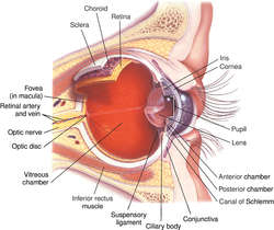
Anatomy
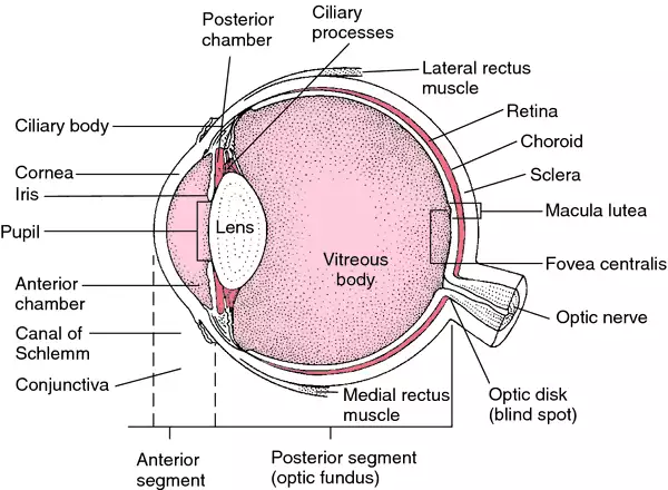
The eyeball has three layers: the inner retina, which contains the photoreceptors; the middle uvea (choroid, ciliary body, and iris); and the outer sclera, which includes the transparent cornea. The eyeball contains two cavities: the anterior cavity and the posterior cavity. The smaller anterior cavity is in front of the lens and is further divided by the iris into an anterior chamber, filled with aqueous humor, and a posterior chamber, filled with the vitreous. Behind the lens is the larger posterior cavity, which contains the vitreous. The lens is behind the iris, held in place by the ciliary body and suspensory ligaments called zonules. The visible portion of the sclera is covered by the conjunctiva. Six extrinsic muscles move the eyeball: the superior, inferior, medial, and lateral rectus muscles, and the superior and inferior oblique muscles.
Nerve supply: The optic (second cranial) nerve contains the fibers from the retina. The eye muscles are supplied by the oculomotor, trochlear, and abducens (third, fourth, and sixth cranial) nerves. The lid muscles are supplied by the facial nerve to the orbicularis oculi and the oculomotor nerve to the levator palpebrae. Sensory fibers to the orbit are furnished by ophthalmic and maxillary fibers of the fifth cranial (trigeminal) nerve. Sympathetic postganglionic fibers originate in the carotid plexus, their cell bodies lying in the superior cervical ganglion. They supply the dilator muscle of the iris. Parasympathetic fibers from the ciliary ganglion pass to the lacrimal gland, ciliary muscle, and constrictor muscles of the iris.
Physiology
Light entering the eye passes through the cornea, then through the pupil, and on through the crystalline lens and the vitreous to the retina. The cornea, aqueous humor, lens, and vitreous are the refracting media of the eye. Changes in the curvature of the lens are brought about by its elasticity and by contraction of the ciliary muscle. These changes focus light rays on the retina, thereby stimulating the rods and cones. The rods detect light, and the cones detect colors in the visible spectrum. The visual area of the cerebral cortex, located in the occipital lobe, registers them as visual sensations. The amount of light entering the eye is regulated by the iris; its constrictor and dilator muscles change the size of the pupil in response to varying amounts of light. The eye can distinguish nearly 8 million differences in color. As the eye ages, objects appear greener. The principal aspects of vision are color sense, light sense, movement, and form sense.
Patient care
When injury to the eye occurs, visual acuity is assessed immediately. If the globe has been penetrated, a suitable eye shield, not an eye patch, is applied. A penetrating foreign body should not be removed. All medications, esp. corticosteroids, are withheld until the patient has been seen by an ophthalmologist.
The patient is assessed for pain and tenderness, redness and discharge, itching, photophobia, increased tearing, blinking, and visual blurring. When any prescribed topical eye medications (drops, ointments, or solutions) are administered, the health care provider should wash his or her hands thoroughly before administering the agent. The patient's head is turned slightly toward the affected eye; his or her cooperation is necessary to keep the eye wide open. Drops are instilled in the conjunctival sac (not on the orb), and pressure is applied to the lacrimal apparatus in the inner canthus if it is necessary to prevent systemic absorption. Ointments are applied along the palpebral border from the inner to the outer canthus, and solutions are instilled from the inner to the outer canthus. Touching the dropper or tip of the medication container to the eye should be avoided, and hands should be washed immediately after the procedure.
Both patient and family are taught correct methods for instilling prescribed medications. Patients with visual defects are protected from injury, and family members are taught safety measures. Patients with insufficient tearing or the inability to blink or close their eyes are protected from corneal injury by applying artificial tears and by gently patching the eyes closed. The importance of periodic eye examinations is emphasized. Persons at risk should protect their eyes from trauma by wearing safety goggles when working with or near dangerous tools or substances. Tinted lenses should be worn to protect the eyes from excessive exposure to bright light. Patients should avoid rubbing their eyes to prevent irritation or possibly infection. See: eyedrops; artificial tears
CAUTION!
Corticosteroids should not be administered topically or systemically until the patient has been seen by a physician, preferably an ophthalmologist.aphakic eye
artificial eye
black eye
Treatment
Application of ice packs during the first 24 hr will inhibit swelling. Hot compresses after the first day may aid absorption of the fluids that produce discoloration.
contact lens–induced red eye
Abbreviation: CLAREcrossed eye
See: cross-eyedark-adapted eye
dominant eye
dry eye
exciting eye
false eye
Artificial eye.fixating eye
glass eye
Artificial eye.Klieg eye
See: Klieg eyelazy eye
Amblyopia.light-adapted eye
squinting eye
sympathizing eye

eye
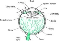
the light-receptor organ of animals. Such organs range in complexity from the single-lensed ocellus found in insects and some other invertebrates, to the vertebrate eye. The vertebrate eye consists of three layers (see Fig. 156 ). The fibrous outer coat consists of the SCLERA and the transparent CORNEA; the middle vascular layer is made up of the CHOROID, the CILIARY BODY and the IRIS; and the RETINA is the inner sensory layer. The two outer layers of the eye serve to support the retina or to focus images of the visual world on the retina.Light enters the eye through the cornea and is refracted through the AQUEOUS HUMOUR on to the lens. The shape of the lens can be altered (see ACCOMMODATION) to focus the light through the VITREOUS HUMOUR on to the retina, where light-receptive ROD CELLS and CONE CELLS (see FOVEA) convert light energy to nerve impulses which pass to the brain via the optic nerve (see BLIND SPOT). An inherited dysfunction of cone cells in some individuals, more common in males than females, can lead to the inability to distinguish between different colours, a condition known as COLOUR BLINDNESS. The position of the eyes at the front of the head provides BINOCULAR VISION, and movement of the eyes within the ORBIT is controlled by extrinsic eye muscles. Several accessory structures, namely the eyelids, LACHRYMAL GLANDS and CONJUNCTIVA, protect the eye from external damage.
eye
amaurotic eye See amaurosis.
amblyopic eye An eye which has amblyopia. Syn. lazy eye (colloquial).
aphakic eye An eye without the crystalline lens.
artificial eye A prosthesis made of glass or plastic which resembles the eye and which is placed in the socket after enucleation. See ocularist; ocular prosthesis.
axial length of the eye See axial length of the eye.
eye bank An organization that collects, evaluates, stores and distributes eyes from donors. The eyes are used for corneal transplants and research. See keratoplasty.
black eye A colloquial term for a swollen or blue-black spot on the skin of the eyelid caused by effusion of blood as a result of a superficial injury in which the skin is not broken. The correct term is ecchymosis of the eyelid. The condition recovers by itself within 2-3 weeks while changing in colour to yellow. Immediately after the injury, application of ice helps minimize the haemorrhage and swelling. See haematoma.
bleary eye A red and watery eye, with a lacklustre appearance. Lack of sleep is a common cause. Syn. blear eye.e. blink See blink.
compound eye The eye of arthropods composed of a variable number of ommatidia. See corneal facet; ommatidium.
crossed eye's See convergent strabismus.
cyclopean eye Imaginary eye located at a point midway between the two eyes. When the two visual fields overlap and the impressions from the two eyes are combined into a single impression, the apparent direction of a fixated object appears in a direction that emanates from the cyclopean eye.
dark-adapted eye An eye that has been in darkness and is sensitive to low illumination. Syn. scotopic eye.
deviating eye The non-fixating eye in strabismus or under heterophoria testing. Syn. squinting eye.
dominant eye The eye that is dominant when ocular dominance exists. See manoptoscope; hole in the card test.
dry eye This term encompasses various tear film disorders ranging from a mild form causing discomfort, which is usually relieved with artificial tears, to the most common form keratoconjunctivitis sicca. See keratoconjunctivitis sicca.
equatorial plane of the eye See equatorial plane.
exciting eye See sympathetic ophthalmia.
fixating eye The eye that is directed towards the object of regard in strabismus. See deviating eye.
glass eye An artificial eye made of glass. See ocularist; ocular prosthesis.
eye impression See eye impression.
lazy eye See amblyopic eye.
eye lens See eyepiece.
light-adapted eye An eye that has been exposed to light and is insensitive to low illumination. Syn. photopic eye. See light adaptation; duplicity theory.
eye movements See eye movements.
eye patch A piece of material or plastic that is worn over the eye when it has been injured or over the socket when it is missing.
phakic eye An eye that contains the crystalline lens. See phakic.
photopic eye See light-adapted eye.
pink eye See contagious conjunctivitis.
eye position Position of the eye in the orbit, maintained by the extraocular muscles. See primary position; secondary position; tertiary position.
pseudophakic eye An eye fitted with an intraocular lens implant. See intraocular lens.
red eye A colloquial term often used for any condition in which the blood vessels of the conjunctiva or ciliary body are congested. Many conditions result in a red eye (e.g. subconjunctival haemorrhage, pterygium, conjunctivitis, episcleritis, corneal abrasion, corneal erosion, ulcerative keratitis, corneal dendritic ulcer, acute iritis, angle-closure glaucoma, orbital cellulitis, and possibly contact lens wear). See ciliary injection; con-junctival injection.
reduced eye A mathematical model of the optical system of the eye. It consists of a single refracting surface with one nodal point, one principal point and one index of refraction. In the first such model, proposed by Listing in 1853, the refracting surface had a power of 68.3 D and was situated 2.34 mm behind the schematic eye's cornea. It had an index of refraction of 1.35, a radius of curvature of 5.124 mm and a length of 20 mm. Donders' reduced eye was even more simplified. It has a power of 66.7 D, a radius of curvature of 5 mm, an index of refraction of 4/3 and anterior and posterior focal lengths of −15 and +20 mm, respectively, with a refracting surface situated 2 mm behind the schematic eye's cornea. Gullstrand's reduced eye has a radius of curvature of 5.7 mm, an index of refraction of 1.33, a power of 61 D with the refracting surface situated 1.35 mm behind the schematic eye's cornea. Emsley's reduced eye has a power of 60 D, an index of refraction of 4/3 and is situated 1.66 mm behind the schematic eye's cornea, with anterior and posterior focal lengths of −16.67 and +22.22 mm, respectively.
schematic eye A model consisting of various spherical surfaces representing the optical system of a normal eye based on the average dimensions (called the constants of the eye) of the human eye. There are many schematic eyes, although the most commonly used is that of Gullstrand. A great deal of variation among authors stemmed from the difficulty in giving an index of refraction that would represent the heterogeneous character of the crystalline lens. Gullstrand in fact proposed two schematic eyes, one which he called the exact schematic eye and the other which he called the simplified schematic eye in which the divergent effect of the posterior corneal surface is ignored and the cornea replaced by an equivalent surface; the crystalline lens is homogeneous and the optical system is free from aberrations.
scotopic eye See dark-adapted eye.
eye shield 1. See occluder.
sighting-dominant eye The eye that is preferred in monocular tasks, such as looking through a telescope or aiming a firearm.
eye socket The bony orbit which contains the eyeball, the muscles, the nerves, the vessels, the orbital fat and the orbital portion of the lacrimal gland.
eye speculum An instrument designed to hold the eyelids apart during surgery. Syn. blepharostat.
squinting eye See deviating eye.
eye stone A small, smooth shell or other object that can be inserted beneath the eyelid to facilitate the removal of a foreign body from the eye.
sympathetic eye The uninjured eye in sympathetic ophthalmia that becomes secondarily affected. Syn. sympathizing eye.
wall eye A colloquial term referring to (1) a white opaque cornea or (2) a divergent strabismus.
watery eye See epiphora.
eye
(ī) [TA]Patient discussion about eye
Q. How to get rid of puffy eyes? After partying all night, I woke up with puffy eyes. I have an important appointment today, how can I get rid of it?
http://www.howtogetridofstuff.com/health/how-to-get-rid-of-puffy-eyes
Q. What age does eye sight stabilizes? I was just wondering at what age does your eye sight usually level off and stop getting worse? Any ideas much appreciated!
It is also the age the eye is fully grown.
Don't worry it will not get much worse maybe about -0,75.
Take care
Q. What Causes Dry Eyes? I have been suffering from eye dryness lately, what causes this situation?
Latest Searches:
ZPLATE - Zovia - Zovant - Zouchlos' - Zoto - Zosyn - Zostrix - zostervirus - zosteroid - zosteriform - zoster - zorubicin - Zortress - ZORprin - Zorbtive - zorbamycin - Zorac - zopolrestat - zopiclone - Zopfius -
- Service manuals - MBI Corp