Medical term:
hydronephrosis
Hydronephrosis
Definition
Description
Causes and symptoms
- painful, burning urine
- cloudy urine
- pain in the back, flank, or groin
- fever, sweats, chills, and generalized discomfort
Diagnosis
Treatment
Alternative treatment
Prognosis
Prevention
Resources
Organizations
Key terms
hydronephrosis
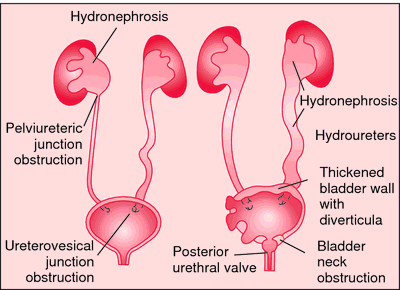
[hi″dro-nĕ-fro´sis]The cause of hydronephrosis is obstruction or atrophy of the urinary tract. Mechanical obstruction may result from ureteral tumors, calculi, benign or malignant hyperplasia of the prostate, or carcinoma of the bladder, urethra, or glans penis. Inflammatory obstruction is the outcome of a urinary tract infection that produces edema and narrowing of the ureters or urethra. Rarely there occurs during pregnancy a loss of muscle tone in the urinary tract. The atony is thought to be induced by placental hormones.
Diagnosis is established by extensive urologic examination with detailed pyelography, which usually reveals the cause of the obstruction and accumulation of fluid in the pelvis.
hy·dro·ne·phro·sis
(hī'drō-ne-frō'sis),hydronephrosis
(hī′drō-nə-frō′sĭs)hydronephrosis
Nephrology Uni- or bilateral expansion of a renal pelvis and calyces, often due to obstructive uropathy which may be linked to HTN and result in permanent renal damage. See Bilateral hydronephrosis, Reflux nephropathy, Unilateral hydrosis.hy·dro·ne·phro·sis
(hī'drō-nĕ-frō'sis)Synonym(s): pelvocaliectasis, uronephrosis.
hydronephrosis
(hi?dro-nef-ro'sis ) [? + nephros, kidney, + osis, condition]
Etiology
Anything that obstructs the ureter or bladder may cause hydronephrosis. Lodged kidney stones are a common cause of unilateral hydronephrosis; bilateral hydronephrosis often results from bladder outlet obstruction (e.g., in men who have hyperplasia of the prostate). Neurogenic bladder dysfunction, pregnancy, urogenital cancer, urinary tract inflammation, congenital malformations, ureteral strictures, and even parasites (schistosomiasis) may cause hydronephrosis. If urinary flow is not restored, the kidney tissue dilates and atrophies, and chronic renal failure may occur.
Symptoms
Hydronephrosis often causes no localizing symptoms, except when it is associated with kidney stones (when the primary symptom is severe flank and abdominal pain that radiates to the groin).
Diagnosis
Ultrasonography of the urinary tract, excretory urography, abdominal CT scan or MRI imaging are used to confirm the diagnosis.
Treatment
Unilateral hydronephrosis caused by a kidney stone resolves spontaneously if the stone passes. If the stone does not pass, procedures such as shock wave lithotripsy, surgical removal of the stone, nephrotomy, or nephrostomy tube drainage of the kidney or placement of a ureteral stent may be needed. Bilateral hydronephrosis caused by prostatic hyperplasia may be relieved if a catheter can be inserted through the obstructed urethra into the bladder, a cystotomy tube is inserted, or the enlarged prostate gland is surgically treated. Hydronephrosis caused by other diseases (e.g., tumors) is often treated with surgical debulking, radiation, or chemotherapy.
Patient care
Renal function studies (e.g., blood urea nitrogen, serum creatinine, and serum potassium levels) are monitored daily. The condition, planned diagnostic procedures, and expected sensations are explained to the patient and family; if the patient is scheduled for a surgical procedure, the surgeon's explanations of the planned procedure are reinforced.
Postoperatively, intake and output, vital signs, and fluid and electrolyte balance are monitored (a rising pulse rate and cool, clammy skin may signal impending hypovolemia or hemorrhage and shock). Prescribed analgesics and noninvasive measures are used to relieve pain as necessary. Postobstructive diuresis may cause the patient to lose great volumes of dilute urine over hours or days along with excessive electrolyte loss. If this occurs, IV fluids are administered at a prescribed constant rate plus an amount equal to a given percentage of the patient's hourly urine output to safely replace intravascular volume. A dietitian can provide assistance to plan a diet consistent with the treatment plan while including foods that the patient enjoys and will eat. If a nephrostomy tube has been inserted, the tube is irrigated as specifically prescribed, checked for patency, and never clamped or allowed to kink. Meticulous skin care is provided to the tube entry site; if urine leaks around the tube, a protective skin barrier is provided to prevent excoriation, and the wound area is bagged to preserve the patient's dignity and to help prevent infection.
If the patient will be discharged with the nephrostomy tube in place, proper care of the tube and skin at the insertion site is taught. Prescribed drug therapies, such as antibiotics, are administered, and the patient is taught about expected outcomes, adverse effects to report, and the necessity to complete the prescribed course of therapy even if feeling better.
hydronephrosis
Ballooning out of the urine collecting system of the kidney, as a result of obstruction to the free outflow of urine at any point below the kidney. This may be due to simple or malignant prostate enlargement, external pressure on, or a stone or blood clot in, a URETER, tumour of the bladder, inflammatory narrowing of the URETHRA or even a very tight foreskin (phimosis). There is pain in the loin, and infection leads to fever and sometimes blood in the urine. Unrelieved hydronephrosis often proceeds to kidney failure.Latest Searches:
zygomaticofrontal - zygomaticofacialis - zygomaticofacial - zygomaticoauricular - zygomatico - zygomatici - zygomatica - zygomatic - zygoma - zygomas - zygodactyly - Zygocotyle - zygion - zygia - zygapophysis - zygapophysiales - zygapophysial - zygapophyseales - zygapophyseal - zygal -
- Service manuals - MBI Corp