Medical term:
immunofluorescence
immunofluorescence
[im″u-no-floo″o-res´ens]a method of determining the location of antigen (or antibody) in a tissue section or smear using a specific antibody (or antigen) labeled with a fluorochrome. There are two major types of immunofluorescence techniques, both based on the antigen--antibody reaction, in which the antibody attaches itself to a specific antigen.
In the direct fluorescent antibody (DFA) method, the antibody coats the antigen, for example, a bacterial cell, and cannot be easily removed by elution (washing). The antibody remains attached to the cell after all nonantibody globulin has been washed away. Since the antibody has been rendered fluorescent by conjugation with fluorescein or another dye, the outline of the bacterial cell that it coats can readily be seen with a special microscope.
In the indirect fluorescent antibody (IFA) method, the specific antibody is allowed to react with the antigen. The nonantibody globulin is then washed off. This is then treated with a labeled antibody to the specific antibody. For example, if the specific antibody was raised in a rabbit, it is then treated with fluorescein-labeled anti-rabbit globulin, which results in a combination of this labeled antibody with the rabbit immunoglobulin already attached to the antigen.
Fluorescent antibody studies have been used in the detection of numerous bacterial, viral, fungal, and protozoan infections and in the identification of many microscopic tissue constituents.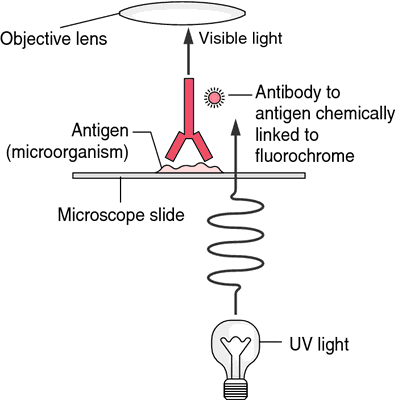

Direct immunofluorescence. In direct immunofluorescence the object is visualized using a fluorescein-tagged antibody. From Hart and Shears, 1997.
Miller-Keane Encyclopedia and Dictionary of Medicine, Nursing, and Allied Health, Seventh Edition. © 2003 by Saunders, an imprint of Elsevier, Inc. All rights reserved.
im·mu·no·fluor·es·cence
(im'yū-nō-flōr-es'ens, i-myū'nō-),An immunohistochemical technique using labeling of antibodies by a fluorescent dye to identify antigenic material specific for the labeled antibody. Specific binding of antibody can be determined microscopically through the production of a characteristic visible light by the application of ultraviolet rays to the preparation.
See also: fluorescent antibody technique.
See also: fluorescent antibody technique.
Farlex Partner Medical Dictionary © Farlex 2012
immunofluorescence
(ĭm′yə-nō-flo͝o-rĕs′əns, -flô-, -flō-)n.
Any of various techniques that use antibodies chemically linked to a fluorescent dye to identify or quantify antigens in a tissue sample.
im′mu·no·fluo·res′cent adj.
The American Heritage® Medical Dictionary Copyright © 2007, 2004 by Houghton Mifflin Company. Published by Houghton Mifflin Company. All rights reserved.
im·mu·no·fluor·es·cence
(im'yū-nō-flōr-es'ĕns)An immunohistochemical technique using labeling of antibodies by fluorescent dyes to identify bacterial, viral, or other antigenic material specific for the labeled antibody; the binding of antibody can be determined microscopically by the application of ultraviolet rays to the preparation.
See also: fluorescent antibody technique
See also: fluorescent antibody technique
Medical Dictionary for the Health Professions and Nursing © Farlex 2012
immunofluorescence
The detection and identification of antigenic material by observing, under the microscope, the fluorescence of known, specific, fluorescein-linked (conjugated) antibodies that have become attached to it.Collins Dictionary of Medicine © Robert M. Youngson 2004, 2005
immunofluorescence
See FLUORESCENT ANTIBODY TECHNIQUE.Collins Dictionary of Biology, 3rd ed. © W. G. Hale, V. A. Saunders, J. P. Margham 2005
Latest Searches:
ZPLATE - Zovia - Zovant - Zouchlos' - Zoto - Zosyn - Zostrix - zostervirus - zosteroid - zosteriform - zoster - zorubicin - Zortress - ZORprin - Zorbtive - zorbamycin - Zorac - zopolrestat - zopiclone - Zopfius -
- Service manuals - MBI Corp