Medical term:
iris
eye
[i]the organ of vision; see also Plates. In the embryo the eye develops as a direct extension of the brain, and thus is a very delicate organ. To protect the eye the bones of the skull are shaped so that an orbital cavity protects the dorsal aspect of each eyeball. In addition, the conjunctival sac covers the front of the eyeball and lines the upper and lower eyelids. Tears from the lacrimal duct constantly wash the eye to remove foreign objects, and the lids and eyelashes help protect the front of the eye.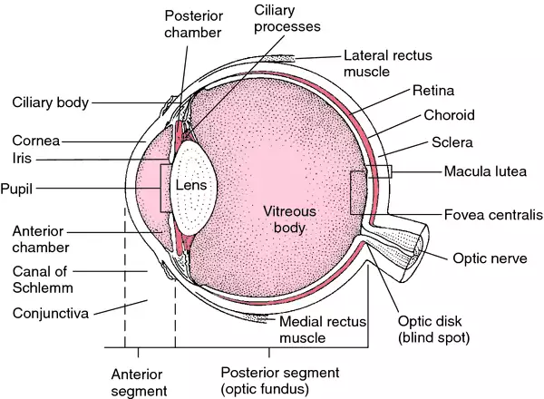
Structure. The eyeball has three coats. The cornea is the clear transparent layer on the front of the eyeball; it is a continuation of the sclera (the white of the eye), the tough outer coat that helps protect the delicate mechanism of the eye. The choroid is the middle layer and contains blood vessels. The third layer, the retina, contains rods and cones, which are specialized cells that are sensitive to light. Behind the cornea and in front of the lens is the iris, the circular pigmented band around the pupil. The iris works much like the diaphragm in a camera, widening or narrowing the pupil to adjust to different light conditions.
Function. (See also vision.) The refraction or bending of light rays so that they focus on the retina and can thus be transmitted to the optic nerve is accomplished by three structures: the aqueous humor, a watery substance between the cornea and lens; the lens, a crystalline structure just behind the iris; and the vitreous humor, a jelly-like substance filling the space between the lens and the retina. Unlike the lens of a camera, the lens of the eye focuses by a process called accommodation. This means that when the eye sees something in the distance, muscles pull the lens, stretching it until it is thin and almost flat, so that the light rays are only slightly bent as they pass through it. When the object is close, the muscles relax and the elastic lens becomes thicker, bending the light rays and focusing them on the retina.ƒ
Because the eye must function under many different circumstances, there are two types of nerve cells in the retina, with different shapes: the cones and the rods. They cover the full range of adaptation to light, the cones being sensitive in bright light, and the rods in dim light. The cones are responsible for color vision. There are three types of cones, each containing a substance that reacts to light of a different color, one set for red, one for green, and one for violet. These are the primary colors in light, which, when mixed together, give white. White light stimulates all three sets of color cells; any other color stimulates one or two.
The optic nerve, which transmits the nerve impulses from the retina to the visual center of the brain, contains nerve fibers from the many nerve cells in the retina. The small spot where it leaves the retina does not have any light-sensitive cells, and is called the blind spot.
The eyes are situated in the front of the head in such a way that human beings have stereoscopic vision, the ability to judge distances. Because the eyes are set apart, each eye sees farther around an object on its own side than does the other. The brain superimposes the two slightly different images and judges distances from the composite image.
Disorders of the Eye. If the eyeball is too short or too long, the lens focuses the image not on the retina but behind or in front of it. The former condition is called hyperopia (or farsightedness) and the latter myopia (or nearsightedness). An irregularity in the curvature of the cornea or lens can cause the impaired vision of astigmatism. strabismus (or squint or crossed eyes) is usually caused by weakness in muscles that control movement of the eyeball. conjunctivitis is an inflammation of the membrane that covers the front of the eyeball and lines the eyelids. When small pieces of the retina become detached from the underlying layers, the result is a retinal detachment; surgery may be necessary to prevent blindness. presbyopia (usually taking the form of hyperopia) occurs in older persons and develops as the lens loses its elasticity with the passing years. Correction is easily made with properly prescribed eyeglasses.ƒ
Foreign bodies in the eyes are common occurrences. Protective eyewear should be worn by individuals at risk. Cinders, grit, or other foreign bodies are best removed by lifting the eyelid by the lashes. The foreign body will usually remain on the surface of the lid, and can easily be removed. Particles embedded in the eyeball must be removed by a qualified health care professional.
Eyestrain is fatigue of the eyes caused by improper use, uncorrected defects in the vision, or an eye disorder. Symptoms may include aching or pains in the eyes, or a hot, scratchy feeling in the eyelids. Headache, blurring or dimness of vision, and sometimes dizziness or nausea may also occur.

Anatomic features of the eye. From Ignatavicius and Workman, 2002.
artificial eye a glass or plastic prosthesis inserted in the eye socket to replace the eyeball; most are designed to be worn day and night. When patients become debilitated and unable to care for such a prosthesis, they must depend on members of the health care team to give proper care according to the chosen preferred routine.ƒ
Cleaning of a prosthetic eye is similar in principle to care of dentures; both are handled with care to avoid damage and are cleansed according to good hygienic principles. The prosthesis is removed while the patient is lying down so that it falls into the hand and is not likely to be dropped and broken. It is removed by depressing the lower eyelid, allowing the prosthesis to slide out and down. Mild soap and water are most often used for cleansing the prosthesis. Alcohol or other chemicals can damage prostheses made of plastic. If it is not replaced in the socket immediately after cleansing, it is stored in water or contact lens soaking solution. Insertion of the prosthesis is done by lifting the upper eyelid with the thumb or forefinger and placing its notched edge toward the nose. It is placed as far as possible under the upper lid and then the lower lid is depressed to allow it to slip into place. The process can be made easier by first moistening the prosthesis with water. If it is necessary to wipe the eye area of a patient wearing a prosthesis, one should gently wipe toward the nose in order not to dislodge the prosthesis.
Cleaning of a prosthetic eye is similar in principle to care of dentures; both are handled with care to avoid damage and are cleansed according to good hygienic principles. The prosthesis is removed while the patient is lying down so that it falls into the hand and is not likely to be dropped and broken. It is removed by depressing the lower eyelid, allowing the prosthesis to slide out and down. Mild soap and water are most often used for cleansing the prosthesis. Alcohol or other chemicals can damage prostheses made of plastic. If it is not replaced in the socket immediately after cleansing, it is stored in water or contact lens soaking solution. Insertion of the prosthesis is done by lifting the upper eyelid with the thumb or forefinger and placing its notched edge toward the nose. It is placed as far as possible under the upper lid and then the lower lid is depressed to allow it to slip into place. The process can be made easier by first moistening the prosthesis with water. If it is necessary to wipe the eye area of a patient wearing a prosthesis, one should gently wipe toward the nose in order not to dislodge the prosthesis.
cross eye esotropia.
dry eye keratoconjunctivitis sicca.
pink eye popular term for acute contagious conjunctivitis.
raccoon e's ecchymotic areas surrounding both eyes, suggestive of a basilar skull fracture.
wall eye exotropia.
Miller-Keane Encyclopedia and Dictionary of Medicine, Nursing, and Allied Health, Seventh Edition. © 2003 by Saunders, an imprint of Elsevier, Inc. All rights reserved.
i·ris
, pl.ir·i·des
(ī'ris, ir'i-dēz), [TA]The anterior division of the vascular tunic of the eye, a diaphragm, perforated in the center (the pupil), attached peripherally to the scleral spur; it is composed of stroma and a double layer of pigmented retinal epithelium from which are derived the sphincter and dilator muscles of the pupil.
Synonym(s): orris
[G. rainbow, the iris of the eye]
Farlex Partner Medical Dictionary © Farlex 2012
iris
(ī′rĭs)n. pl. irises or irides (ī′rĭ-dēz′, ĭr′ĭ-)
1. The pigmented, round, contractile membrane of the eye, suspended between the cornea and lens and perforated by the pupil. It regulates the amount of light entering the eye.
2. Any of numerous widely cultivated plants of the genus Iris, having narrow sword-shaped leaves and showy, variously colored flowers.
3. A rainbow or rainbowlike display of colors.
4. An iris diaphragm.
The American Heritage® Medical Dictionary Copyright © 2007, 2004 by Houghton Mifflin Company. Published by Houghton Mifflin Company. All rights reserved.
iris
Fringe medicineAn essence which, in the pseudoscience of flower essence therapy, is believed to provide artisitic vision radiance and perspective.
Segen's Medical Dictionary. © 2012 Farlex, Inc. All rights reserved.
i·ris
, pl. irides (īris, iri-dēz) [TA]The anterior division of the vascular tunic of the eye, a diaphragm, perforated in the center (the pupil), attached peripherally to the scleral spur; it is composed of stroma and a double layer of pigmented retinal epithelium from which are derived the sphincter and dilator muscles of the pupil.
[G. rainbow, the iris of the eye]
Medical Dictionary for the Health Professions and Nursing © Farlex 2012
iris
The coloured diaphragm of the eye forming the rear wall of the front, water-filled, chamber and lying immediately in front of the CRYSTALLINE LENS. The iris has a central opening, the pupil. It contains circular muscle fibres to constrict the pupil and radial fibres to enlarge (dilate) it.Collins Dictionary of Medicine © Robert M. Youngson 2004, 2005
iris
the pigmented part of the vertebrate eye. It consists of a thin sheet of tissue, attached at its outer edge to the CILIARY BODY, which has radiating muscles which can increase the size of the central pupil and a central ring of muscle around the pupil which, on contraction, causes a decrease in its size. The iris thus regulates the amount of light entering the eye. See ALBINISM for photophobia.Collins Dictionary of Biology, 3rd ed. © W. G. Hale, V. A. Saunders, J. P. Margham 2005
Iris (plural, irides)
The circular pigmented membrane behind the cornea of the eye that gives the eye its color. The iris surrounds a central opening called the pupil.
Mentioned in: Eye Cancer, Eye Examination, Glaucoma, Hyperopia, Radial Keratotomy, Retinal Vein Occlusion, Trabeculectomy, Uveitis, Vitiligo
Gale Encyclopedia of Medicine. Copyright 2008 The Gale Group, Inc. All rights reserved.
iris
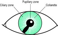
The anterior part of the vascular tunic of the eye, which is situated in front of the crystalline lens and behind the cornea. It has the shape of a circular membrane with a perforation in the centre (the pupil) and is attached peripherally to the ciliary body. The iris forms a curtain dividing the space between the cornea and the lens into the anterior and posterior chambers of the eye. The anterior surface of the iris is divided into two portions: the largest peripheral ciliary zone and the inner pupillary zone. The two zones are separated by a zigzag line, the collarette. The iris consists of four layers which are, starting in the front: (1) the layer of fibrocytes and melanocytes; (2) the stroma in which are embedded the following structures: (a) the sphincter pupillae muscle which constricts the pupil and is supplied mainly by parasympathetic fibres via the third cranial nerve, (b) the vessels which form the bulk of the iris, and (c) the pigment cells; (3) the posterior membrane consisting of plain muscle fibres which constitute the dilator muscle which is supplied mainly by sympathetic motor fibres, via the long ciliary nerves; (4) the posterior epithelium which is highly pigmented.Sensory fibres from the iris are contained in the nasociliary branch of the ophthalmic nerve. The blood supply is provided by the ciliary arteries. The colour of the iris is blue in babies belonging to the white races and changes colour after a few months of life as pigment is deposited in the anterior limiting layer and the stroma. Iris colour is inherited; brown as a dominant trait and blue as a recessive trait. Iris patterns are unique for each individual and can be used as a type of identification. The function of the iris and pupil is to regulate the amount of light admitted into the eye, to optimize the depth of focus and to mitigate ocular aberrations.See cell, clump; corectopia; Fig. C 13; Fuchs, crypts of; heterochromia; inheritance; iridectomy; iridodialysis; iridology; iritis; melanin; membrane, pupillary; polycoria; reflex, pupil light.

| Table I6 Differential diagnosis* between acute conjunctivitis, acute iritis and angle-closure glaucoma | ||||||
| acute conjunctivitis | acute iritis (anterior uveitis) | angle-closure glaucoma | ||||
| signs | ||||||
| injection | conjunctival | ciliary | conjunctival and ciliary | |||
| pupil | normal | contracted | semi-dilated and fixed | |||
| intraocular pressure | normal | normal or low, occasionally increased | high | |||
| cornea | normal | KP | Oedematous | |||
| anterior chamber | normal depth | normal depth, aqueous flare | shallow | |||
| iris | normal | faded | faded | |||
| view of fundus | clear | misty | almost invisible | |||
| symptoms | ||||||
| pain | irritation | moderate to servere | very severe and radiating | |||
| photophobia | slight | marked | slight | |||
| lacrimation | watery, purulent or mucopurulent | watery | watery | |||
| vision | normal | slightly reduced | much reduced, haloes | |||
| onset | gradual | rapid | sudden | |||
| systemic complications | none | malaise or fever | nausea and vomiting | |||
| *This is a guide, as individual cases vary according to the cause and severity of the disease. | ||||||

Fig. C13 Coloboma of the iris
Millodot: Dictionary of Optometry and Visual Science, 7th edition. © 2009 Butterworth-Heinemann
i·ris
, pl. irides (īris, iri-dēz) [TA]The anterior division of the vascular tunic of the eye, a diaphragm, perforated in the center (the pupil), attached peripherally to the scleral spur.
[G. rainbow, the iris of the eye]
Medical Dictionary for the Dental Professions © Farlex 2012
eye
[i]the organ of vision; see also Plates. In the embryo the eye develops as a direct extension of the brain, and thus is a very delicate organ. To protect the eye the bones of the skull are shaped so that an orbital cavity protects the dorsal aspect of each eyeball. In addition, the conjunctival sac covers the front of the eyeball and lines the upper and lower eyelids. Tears from the lacrimal duct constantly wash the eye to remove foreign objects, and the lids and eyelashes help protect the front of the eye.
Structure. The eyeball has three coats. The cornea is the clear transparent layer on the front of the eyeball; it is a continuation of the sclera (the white of the eye), the tough outer coat that helps protect the delicate mechanism of the eye. The choroid is the middle layer and contains blood vessels. The third layer, the retina, contains rods and cones, which are specialized cells that are sensitive to light. Behind the cornea and in front of the lens is the iris, the circular pigmented band around the pupil. The iris works much like the diaphragm in a camera, widening or narrowing the pupil to adjust to different light conditions.
Function. (See also vision.) The refraction or bending of light rays so that they focus on the retina and can thus be transmitted to the optic nerve is accomplished by three structures: the aqueous humor, a watery substance between the cornea and lens; the lens, a crystalline structure just behind the iris; and the vitreous humor, a jelly-like substance filling the space between the lens and the retina. Unlike the lens of a camera, the lens of the eye focuses by a process called accommodation. This means that when the eye sees something in the distance, muscles pull the lens, stretching it until it is thin and almost flat, so that the light rays are only slightly bent as they pass through it. When the object is close, the muscles relax and the elastic lens becomes thicker, bending the light rays and focusing them on the retina.ƒ
Because the eye must function under many different circumstances, there are two types of nerve cells in the retina, with different shapes: the cones and the rods. They cover the full range of adaptation to light, the cones being sensitive in bright light, and the rods in dim light. The cones are responsible for color vision. There are three types of cones, each containing a substance that reacts to light of a different color, one set for red, one for green, and one for violet. These are the primary colors in light, which, when mixed together, give white. White light stimulates all three sets of color cells; any other color stimulates one or two.
The optic nerve, which transmits the nerve impulses from the retina to the visual center of the brain, contains nerve fibers from the many nerve cells in the retina. The small spot where it leaves the retina does not have any light-sensitive cells, and is called the blind spot.
The eyes are situated in the front of the head in such a way that human beings have stereoscopic vision, the ability to judge distances. Because the eyes are set apart, each eye sees farther around an object on its own side than does the other. The brain superimposes the two slightly different images and judges distances from the composite image.
Because the eye must function under many different circumstances, there are two types of nerve cells in the retina, with different shapes: the cones and the rods. They cover the full range of adaptation to light, the cones being sensitive in bright light, and the rods in dim light. The cones are responsible for color vision. There are three types of cones, each containing a substance that reacts to light of a different color, one set for red, one for green, and one for violet. These are the primary colors in light, which, when mixed together, give white. White light stimulates all three sets of color cells; any other color stimulates one or two.
The optic nerve, which transmits the nerve impulses from the retina to the visual center of the brain, contains nerve fibers from the many nerve cells in the retina. The small spot where it leaves the retina does not have any light-sensitive cells, and is called the blind spot.
The eyes are situated in the front of the head in such a way that human beings have stereoscopic vision, the ability to judge distances. Because the eyes are set apart, each eye sees farther around an object on its own side than does the other. The brain superimposes the two slightly different images and judges distances from the composite image.
Disorders of the Eye. If the eyeball is too short or too long, the lens focuses the image not on the retina but behind or in front of it. The former condition is called hyperopia (or farsightedness) and the latter myopia (or nearsightedness). An irregularity in the curvature of the cornea or lens can cause the impaired vision of astigmatism. strabismus (or squint or crossed eyes) is usually caused by weakness in muscles that control movement of the eyeball. conjunctivitis is an inflammation of the membrane that covers the front of the eyeball and lines the eyelids. When small pieces of the retina become detached from the underlying layers, the result is a retinal detachment; surgery may be necessary to prevent blindness. presbyopia (usually taking the form of hyperopia) occurs in older persons and develops as the lens loses its elasticity with the passing years. Correction is easily made with properly prescribed eyeglasses.ƒ
Foreign bodies in the eyes are common occurrences. Protective eyewear should be worn by individuals at risk. Cinders, grit, or other foreign bodies are best removed by lifting the eyelid by the lashes. The foreign body will usually remain on the surface of the lid, and can easily be removed. Particles embedded in the eyeball must be removed by a qualified health care professional.
Eyestrain is fatigue of the eyes caused by improper use, uncorrected defects in the vision, or an eye disorder. Symptoms may include aching or pains in the eyes, or a hot, scratchy feeling in the eyelids. Headache, blurring or dimness of vision, and sometimes dizziness or nausea may also occur.
Foreign bodies in the eyes are common occurrences. Protective eyewear should be worn by individuals at risk. Cinders, grit, or other foreign bodies are best removed by lifting the eyelid by the lashes. The foreign body will usually remain on the surface of the lid, and can easily be removed. Particles embedded in the eyeball must be removed by a qualified health care professional.
Eyestrain is fatigue of the eyes caused by improper use, uncorrected defects in the vision, or an eye disorder. Symptoms may include aching or pains in the eyes, or a hot, scratchy feeling in the eyelids. Headache, blurring or dimness of vision, and sometimes dizziness or nausea may also occur.
Anatomic features of the eye. From Ignatavicius and Workman, 2002.
artificial eye a glass or plastic prosthesis inserted in the eye socket to replace the eyeball; most are designed to be worn day and night. When patients become debilitated and unable to care for such a prosthesis, they must depend on members of the health care team to give proper care according to the chosen preferred routine.ƒ
Cleaning of a prosthetic eye is similar in principle to care of dentures; both are handled with care to avoid damage and are cleansed according to good hygienic principles. The prosthesis is removed while the patient is lying down so that it falls into the hand and is not likely to be dropped and broken. It is removed by depressing the lower eyelid, allowing the prosthesis to slide out and down. Mild soap and water are most often used for cleansing the prosthesis. Alcohol or other chemicals can damage prostheses made of plastic. If it is not replaced in the socket immediately after cleansing, it is stored in water or contact lens soaking solution. Insertion of the prosthesis is done by lifting the upper eyelid with the thumb or forefinger and placing its notched edge toward the nose. It is placed as far as possible under the upper lid and then the lower lid is depressed to allow it to slip into place. The process can be made easier by first moistening the prosthesis with water. If it is necessary to wipe the eye area of a patient wearing a prosthesis, one should gently wipe toward the nose in order not to dislodge the prosthesis.
Cleaning of a prosthetic eye is similar in principle to care of dentures; both are handled with care to avoid damage and are cleansed according to good hygienic principles. The prosthesis is removed while the patient is lying down so that it falls into the hand and is not likely to be dropped and broken. It is removed by depressing the lower eyelid, allowing the prosthesis to slide out and down. Mild soap and water are most often used for cleansing the prosthesis. Alcohol or other chemicals can damage prostheses made of plastic. If it is not replaced in the socket immediately after cleansing, it is stored in water or contact lens soaking solution. Insertion of the prosthesis is done by lifting the upper eyelid with the thumb or forefinger and placing its notched edge toward the nose. It is placed as far as possible under the upper lid and then the lower lid is depressed to allow it to slip into place. The process can be made easier by first moistening the prosthesis with water. If it is necessary to wipe the eye area of a patient wearing a prosthesis, one should gently wipe toward the nose in order not to dislodge the prosthesis.
cross eye esotropia.
dry eye keratoconjunctivitis sicca.
pink eye popular term for acute contagious conjunctivitis.
raccoon e's ecchymotic areas surrounding both eyes, suggestive of a basilar skull fracture.
wall eye exotropia.
Miller-Keane Encyclopedia and Dictionary of Medicine, Nursing, and Allied Health, Seventh Edition. © 2003 by Saunders, an imprint of Elsevier, Inc. All rights reserved.
i·ris
, pl.ir·i·des
(ī'ris, ir'i-dēz), [TA]The anterior division of the vascular tunic of the eye, a diaphragm, perforated in the center (the pupil), attached peripherally to the scleral spur; it is composed of stroma and a double layer of pigmented retinal epithelium from which are derived the sphincter and dilator muscles of the pupil.
Synonym(s): orris
[G. rainbow, the iris of the eye]
Farlex Partner Medical Dictionary © Farlex 2012
iris
(ī′rĭs)n. pl. irises or irides (ī′rĭ-dēz′, ĭr′ĭ-)
1. The pigmented, round, contractile membrane of the eye, suspended between the cornea and lens and perforated by the pupil. It regulates the amount of light entering the eye.
2. Any of numerous widely cultivated plants of the genus Iris, having narrow sword-shaped leaves and showy, variously colored flowers.
3. A rainbow or rainbowlike display of colors.
4. An iris diaphragm.
The American Heritage® Medical Dictionary Copyright © 2007, 2004 by Houghton Mifflin Company. Published by Houghton Mifflin Company. All rights reserved.
iris
Fringe medicineAn essence which, in the pseudoscience of flower essence therapy, is believed to provide artisitic vision radiance and perspective.
Segen's Medical Dictionary. © 2012 Farlex, Inc. All rights reserved.
i·ris
, pl. irides (īris, iri-dēz) [TA]The anterior division of the vascular tunic of the eye, a diaphragm, perforated in the center (the pupil), attached peripherally to the scleral spur; it is composed of stroma and a double layer of pigmented retinal epithelium from which are derived the sphincter and dilator muscles of the pupil.
[G. rainbow, the iris of the eye]
Medical Dictionary for the Health Professions and Nursing © Farlex 2012
iris
The coloured diaphragm of the eye forming the rear wall of the front, water-filled, chamber and lying immediately in front of the CRYSTALLINE LENS. The iris has a central opening, the pupil. It contains circular muscle fibres to constrict the pupil and radial fibres to enlarge (dilate) it.Collins Dictionary of Medicine © Robert M. Youngson 2004, 2005
iris
the pigmented part of the vertebrate eye. It consists of a thin sheet of tissue, attached at its outer edge to the CILIARY BODY, which has radiating muscles which can increase the size of the central pupil and a central ring of muscle around the pupil which, on contraction, causes a decrease in its size. The iris thus regulates the amount of light entering the eye. See ALBINISM for photophobia.Collins Dictionary of Biology, 3rd ed. © W. G. Hale, V. A. Saunders, J. P. Margham 2005
Iris (plural, irides)
The circular pigmented membrane behind the cornea of the eye that gives the eye its color. The iris surrounds a central opening called the pupil.
Mentioned in: Eye Cancer, Eye Examination, Glaucoma, Hyperopia, Radial Keratotomy, Retinal Vein Occlusion, Trabeculectomy, Uveitis, Vitiligo
Gale Encyclopedia of Medicine. Copyright 2008 The Gale Group, Inc. All rights reserved.
iris
The anterior part of the vascular tunic of the eye, which is situated in front of the crystalline lens and behind the cornea. It has the shape of a circular membrane with a perforation in the centre (the pupil) and is attached peripherally to the ciliary body. The iris forms a curtain dividing the space between the cornea and the lens into the anterior and posterior chambers of the eye. The anterior surface of the iris is divided into two portions: the largest peripheral ciliary zone and the inner pupillary zone. The two zones are separated by a zigzag line, the collarette. The iris consists of four layers which are, starting in the front: (1) the layer of fibrocytes and melanocytes; (2) the stroma in which are embedded the following structures: (a) the sphincter pupillae muscle which constricts the pupil and is supplied mainly by parasympathetic fibres via the third cranial nerve, (b) the vessels which form the bulk of the iris, and (c) the pigment cells; (3) the posterior membrane consisting of plain muscle fibres which constitute the dilator muscle which is supplied mainly by sympathetic motor fibres, via the long ciliary nerves; (4) the posterior epithelium which is highly pigmented.Sensory fibres from the iris are contained in the nasociliary branch of the ophthalmic nerve. The blood supply is provided by the ciliary arteries. The colour of the iris is blue in babies belonging to the white races and changes colour after a few months of life as pigment is deposited in the anterior limiting layer and the stroma. Iris colour is inherited; brown as a dominant trait and blue as a recessive trait. Iris patterns are unique for each individual and can be used as a type of identification. The function of the iris and pupil is to regulate the amount of light admitted into the eye, to optimize the depth of focus and to mitigate ocular aberrations.See cell, clump; corectopia; Fig. C 13; Fuchs, crypts of; heterochromia; inheritance; iridectomy; iridodialysis; iridology; iritis; melanin; membrane, pupillary; polycoria; reflex, pupil light.
| Table I6 Differential diagnosis* between acute conjunctivitis, acute iritis and angle-closure glaucoma | ||||||
| acute conjunctivitis | acute iritis (anterior uveitis) | angle-closure glaucoma | ||||
| signs | ||||||
| injection | conjunctival | ciliary | conjunctival and ciliary | |||
| pupil | normal | contracted | semi-dilated and fixed | |||
| intraocular pressure | normal | normal or low, occasionally increased | high | |||
| cornea | normal | KP | Oedematous | |||
| anterior chamber | normal depth | normal depth, aqueous flare | shallow | |||
| iris | normal | faded | faded | |||
| view of fundus | clear | misty | almost invisible | |||
| symptoms | ||||||
| pain | irritation | moderate to servere | very severe and radiating | |||
| photophobia | slight | marked | slight | |||
| lacrimation | watery, purulent or mucopurulent | watery | watery | |||
| vision | normal | slightly reduced | much reduced, haloes | |||
| onset | gradual | rapid | sudden | |||
| systemic complications | none | malaise or fever | nausea and vomiting | |||
| *This is a guide, as individual cases vary according to the cause and severity of the disease. | ||||||
Fig. C13 Coloboma of the iris
Millodot: Dictionary of Optometry and Visual Science, 7th edition. © 2009 Butterworth-Heinemann
i·ris
, pl. irides (īris, iri-dēz) [TA]The anterior division of the vascular tunic of the eye, a diaphragm, perforated in the center (the pupil), attached peripherally to the scleral spur.
[G. rainbow, the iris of the eye]
Medical Dictionary for the Dental Professions © Farlex 2012
Latest Searches:
zygomaticofrontal - zygomaticofacialis - zygomaticofacial - zygomaticoauricular - zygomatico - zygomatici - zygomatica - zygomatic - zygoma - zygomas - zygodactyly - Zygocotyle - zygion - zygia - zygapophysis - zygapophysiales - zygapophysial - zygapophyseales - zygapophyseal - zygal -
- Service manuals - MBI Corp