retina
[ret´ĭ-nah] the innermost of the three tunics of the
eye, surrounding the vitreous body and continuous posteriorly with the optic nerve. The retina is composed of light-sensitive neurons arranged in three layers; the first layer is made up of rods and cones and the other two transmit impulses from the rods and cones to the
optic nerve. The rods are sensitive in dim light, and the cones are sensitive in bright light and are responsible for color vision.
Disorders of the Retina. retinopathies are pathologic conditions of the retina; they occur in conjunction with certain systemic disorders, such as hypertension, nephritis, toxemia of pregnancy, and diabetes mellitus.
detachment of the retina is complete or partial separation of the retina from the choroid, the middle coat of the eyeball. It occurs most often in persons with myopia (nearsightedness), but it also can result from trauma to the head.
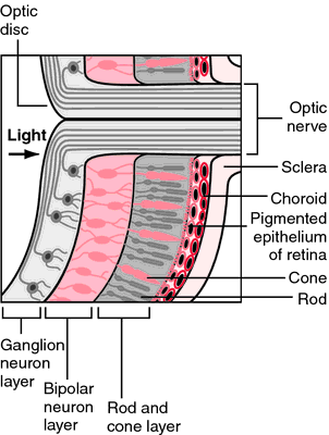
Structure of the retina showing the rods and cones. From Applegate, 2000.
Miller-Keane Encyclopedia and Dictionary of Medicine, Nursing, and Allied Health, Seventh Edition. © 2003 by Saunders, an imprint of Elsevier, Inc. All rights reserved.
ret·i·na
(ret'i-nă), [TA] Grossly, the retina consists of three parts: optic, ciliary, and iridial. The optic part, the physiologic portion that receives the visual light rays, is further divided into two parts, the pigmented part (pigment epithelium) and the nervous part, which are arranged in the following ten layers: pigmented layer; layer of inner and outer segments (of rods and cones); outer limiting layer (actually a row of junctional complexes); outer nuclear layer; outer plexiform layer; inner nuclear layer; inner plexiform layer; ganglionic (cell) layer; layer of nerve fibers; and inner limiting layer. Layers 2-10 comprise the neural layer. At the posterior pole of the visual axis is the macula, in the center of which is the fovea, the area of acute vision. Here, layers 6-9 and blood vessels are absent, and only elongated cones are present. About 3 mm medial to the fovea is the optic disc, where axons of the ganglionic cells converge to form the optic nerve. The ciliary and iridial parts of the retina are forward prolongations of the pigmented layer and a layer of supporting columnar or epithelial cells over the ciliary body and the posterior surface of the iris, respectively.
Synonym(s): optomeninx
[Mediev. L. prob. fr. L. rete, a net]
Farlex Partner Medical Dictionary © Farlex 2012
retina
(rĕt′n-ə)n. pl. retinas or
retinae (rĕt′n-ē′) A delicate, multilayered, light-sensitive membrane lining the inner eyeball and connected by the optic nerve to the brain.
ret′i·nal adj.
The American Heritage® Medical Dictionary Copyright © 2007, 2004 by Houghton Mifflin Company. Published by Houghton Mifflin Company. All rights reserved.
ret·i·na
(ret'i-nă) [TA] The light-sensitive membrane forming the innermost layer of the eyeball. Grossly, the retina consists of three parts: optic part of retina, ciliary part of retina, and iridial part of retina. The optic part, the physiologic portion that receives the visual light rays, is further divided into two parts, pigmented part (pigment epithelium) and nervous part, which are arranged in the following layers: 1) pigment epithelium; 2) layer of rods and cones; 3) external limiting lamina, actually a row of junctional complexes; 4) external nuclear lamina; 5) external plexiform lamina; 6) internal nuclear lamina; 7) internal plexiform lamina; 8) ganglionic cell lamina; 9) lamina of nerve fibers; 10) internal limiting lamina. Layers 2-10 comprise the nervous part. At the posterior pole of the visual axis is the macula, in the center of which is the fovea, the area of acute vision. Here layers 6-9 and blood vessels are absent, and only elongated cones are present. About 3 mm medial to the fovea is the optic disc, where axons of the ganglionic cells converge to form the optic nerve. The ciliary and iridial parts of the retina are forward prolongations of the pigmented layer and a layer of supporting columnar or epithelial cells over the ciliary body and the posterior surface of the iris, respectively.
[Mediev. L. prob. fr. L. rete, a net]
Medical Dictionary for the Health Professions and Nursing © Farlex 2012
retina
(rĕt′ĭ-nă) plural.retinae [L.] 

RETINA OF THE RIGHT EYE
RETINA: Microscopic structure of optic disk area
The innermost layer of the eye, which receives images transmitted through the lens and contains the receptors for vision, the rods and cones. See:
illustrationretinal (-năl),
adjectiveThe retina is a light-sensitive membrane on which light rays are focused. It extends from the entrance point of the optic nerve anteriorly to the margin of the pupil, completely lining the interior of the eye. It consists of three parts. The pars optica, the nervous or sensory portion, extends from the optic disk forward to the ora serrata, a wavy line immediately behind the ciliary process; the pars ciliaris lines the inner surface of the ciliary process; and the pars iridica forms the posterior surface of the iris. Slightly lateral to the posterior pole of the eye is a small, oval, yellowish spot, the macula lutea, in the center of which is a depression, the fovea centralis. This region contains only cones and is the region of the most acute vision. About 3.5 mm nasally from the fovea is the optic papilla (optic disk), where nerve fibers from the retina make their exit and form the optic nerve. This region is devoid of rods and cones and is insensitive to light; hence it is named the blind spot.
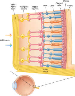
The layers of the retina, in the order light strikes them, are the optic nerve fiber layer, ganglion cell layer, inner synaptic layer, bipolar cell layer, outer synaptic layer, layer of rods and cones, and pigment epithelium. See: illustration
Color
The retina is normally red, reflecting blood flow, and is pale in anemia or ischemia.
Vessels
The arteries are branches of a single central artery, which is a branch of the ophthalmic artery. The central artery enters at the center of the optic papilla and supplies the inner layers of the retina. The outer layers, including rods and cones, are nourished by capillaries of the choroid layer. The veins lack muscular coats. They parallel the arteries; blood leaves by a central vein that leads to the superior ophthalmic vein.
coarctate retina
A condition in which there is an effusion of fluid between the retina and choroid, giving the retina a funnel shape.
shot-silk retina
A retina having an opalescent appearance, sometimes seen in young persons.
tigroid retina
A retina having a spotted or striped appearance, seen in retinitis pigmentosa.
Medical Dictionary, © 2009 Farlex and Partners
retina
The complex membranous network of nerve cells, fibres and photoreceptors that lines the inside of the back of the eye and converts optical images formed by the lens system of the eye into nerve impulses. The retina contains colour-blind but very sensitive rods and colour-sensitive cones and a computing system that refines the signals produced by these. The impulses leave each retina by way of about one million nerve fibres that form the optic nerve.Collins Dictionary of Medicine © Robert M. Youngson 2004, 2005
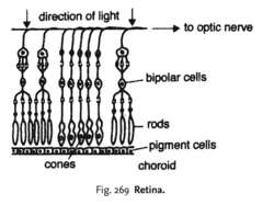
Fig. 269 Retina.
retina
the lining of the interior of the vertebrate eye containing a concentration of photoreceptor cells known as rods and cones that are connected to the optic nerve via BIPOLAR CELLS. The retina lies immediately in front of the vascular choroid layer which nourishes it.Collins Dictionary of Biology, 3rd ed. © W. G. Hale, V. A. Saunders, J. P. Margham 2005
Retina
Light sensitive layer of the eye, that consists of four major layers: the outer neural layer, containing nerve cells and blood vessels, the photoreceptor layer, a single layer that contains the light sensing rods and cones, the pigmented retinal epithelium (PRE) and the choroid, consisting of connective tissue and capillaries.
Mentioned in: Cataract Surgery, Cataracts, Color Blindness, Eye and Orbit Ultrasounds, Eye Examination, Eye Glasses and Contact Lenses, Eye Muscle Surgery, Glaucoma, Hyperopia, Macular Degeneration, Microphthalmia and Anophthalmia, Myopia, Photorefractive Keratectomy and Laser-Assisted In-Situ Keratomileusis, Radial Keratotomy, Retinal Artery Occlusion, Retinal Vein Occlusion, Retinoblastoma, Uveitis, Vitiligo
Gale Encyclopedia of Medicine. Copyright 2008 The Gale Group, Inc. All rights reserved.
retina
The light-receptive, innermost nervous tunic of the eye. It is a thin transparent membrane (about 125 μm near the ora serrata, 350 μm near the macula and 560 μm near the optic disc). The retina proper has an area of about 266 mm
2. It lies between the vitreous body and the choroid, and extends from the optic disc to the ora serrata. Near the posterior pole and temporal to the optic disc is the macula, at the centre of which is the foveola which provides the best visual acuity. The retina contains at least 10 distinct layers, of which there are two synaptic layers. They are from the outermost layer to the innermost: (1) the pigment epithelium; (2) the layer of rods and cones; (3) the external limiting membrane; (4) the outer nuclear layer; (5) the outer molecular (outer plexiform) layer; (6) the inner nuclear layer (which contains the bipolar, amacrine and horizontal cells and nuclei of the fibres of Mueller); (7) the inner molecular (inner plexiform) layer; (8) the ganglion cell layer; (9) the nerve fibre (layer stratum opticum); and (10) the internal limiting membrane. The two synaptic layers where visual signals must synapse as they emerge from the rods and cones on their way to the optic nerve are the two molecular layers (5 and 7) (Fig. R9). The blood supply to the retina is composed of the capillaries from the central retinal artery, which supply the inner two-thirds of the retina up to the outer plexiform layer, and the choriocapillaris, which supplies the outer one-third. There is no retinal circulation in the foveola (avascular zone). A
blood-retina barrier is created by the walls of the retinal capillaries which restrict the movement of molecules, which could be damaging to neural tissue or interfere with function, from the inside to the outside of the capillaries. The blood-retina barrier in the outer third of the retina is formed by the tight junctions of the retinal pigment epithelium cells.
See astrocytes; optic cup; optic disc; ectoderm; ocular fundus;
fibre layer of Henle; macula; neurotransmitter;
neurosensory retina; retinitis; retinopathy; rhodopsin; transduction.
converse retina See inverted retina.
fleck retina Term referring to a retina with multiple, small or yellow spots, which are seen in various conditions: actinic keratopathy, drusen, fundus albipunctatus, fundus flavimaculatus.
inverted retina Term referring to the fact that the retina of vertebrates is orientated so that the light has to pass through all the neuronal layers before reaching the photo-receptors. However, the retina of invertebrates is normally orientated so that light passes first through the photoreceptors as it traverses the retina: such a retina is called a
verted or
converse retina.
lattice degeneration of the retina A vitreoretinal degeneration usually found between the equator and the ora serrata leading to a thinning of the retina and characterized by a lesion made up of fine white lines and some pigmentation. It may result in holes or tears and in rhegmatogenous retinal detachment by which time the patient usually complains of floaters. The condition is most common in myopes and often found in patients with Marfan's syndrome.
See retinal break; retinal detachment; retinoschisis; Marfan's syndrome.
leopard retina See leopard fundus.
neurosensory retina This is composed of all the layers of the retina, except the outer pigmented layer (called retinal pigment epithelium). It comprises three main groups of neurons: (1) the photoreceptors, (2) the bipolar cells, and (3) the ganglion cells. In addition, there are other connecting neurons: the horizontal and amacrine cells. The neurosensory layer is derived embryologically from the inner layer of the optic cup whereas the pigmented layer is derived from the outer layer of the optic cup and they are separated by a potential space which facilitates their separation, as occurs in detached retina.
Syn. sensory retina.
tessellated retina See tessellated fundus.
tigroid retina See tessellated fundus.
verted retina See inverted retina.
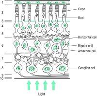
Fig. R9 Schematic representation of the cells and layers of the central primate retina (1: retinal pigment epithelium; 2: layer of rods and cones; 3: external limiting membrane; 4: outer nuclear layer; 5: outer plexiform layer; 6: inner nuclear layer; 7: inner plexiform layer; 8: ganglion cell layer; 9: nerve fibre layer; 10: internal limiting membrane)
| Table R2 Some approximate retinal dimensions |
| | diameter | | |
| structure | | mm | | degrees | | distance from centre of foveola |
| foveola | | 0.4 | | 1.3 | | |
| fovea centralis | | 1.5 | | 5.0 | | |
| macula lutea | | 4.0 | | 14 | | |
| optic disc* | | | | | | |
| horizontal | | 1.8 | | 6.0 | | |
| vertical | | 2.1 | | 7.5 | | |
| nasal disc margin | | | | | | 5.5 mm or 18.5 |
| temporal disc margin | | | | | | 3.5 mm or 12.5 |
| centre of disc | | | | | | 4.6 mm or 15.5 |
| *The figures given for the size of the disc are those corresponding to the blind spot. Anatomically the optic disc is slightly smaller. |
Millodot: Dictionary of Optometry and Visual Science, 7th edition. © 2009 Butterworth-Heinemann
ret·i·na
(ret'i-nă) [TA] Grossly, section of eye that consists of three parts: optic, ciliary, and iridial. The optic part, the physiologic portion that receives the visual light rays, is further divided into two parts, the pigmented part (pigment epithelium) and the nervous part.
[Mediev. L. prob. fr. L. rete, a net]
Medical Dictionary for the Dental Professions © Farlex 2012


