Medical term:
stratigraphy
tomography
[to-mog´rah-fe]any method that produces images of single tissue planes. In conventional radiology, tomographic images (body section radiographs) are produced by motion of the x-ray tube and film or by motion of the patient that blurs the image except in a single plane. In reconstruction tomography (CT and PET) the image is produced by a computer program.
computed tomography (CT) (computerized axial tomography (CAT)) a radiologic imaging modality that uses computer processing to generate an image (CAT scan) of the tissue density in a “slice” as thin as 1 to 10 mm in thickness through the patient's body. These images are spaced at intervals of 0.5 to 1 cm. Cross-sectional anatomy can be reconstructed in several planes without exposing the patient to additional radiation.
Since its introduction in 1972, the use of this modality has grown rapidly. Because it is noninvasive and has high contrast resolution, it has replaced some radiographic procedures using contrast media. It also has a better spatial resolution than scintillation imaging (about 1 mm for CAT compared to 15 mm for a scintillation camera).
A CAT scan is divided into a square matrix of pixels (picture elements). The newer CAT scanners use a high resolution matrix with 256 × 256 or 512 × 512 pixels. The region of the tissue slice corresponding to a pixel has a cross-sectional area of 1 × 1 mm to 2 × 2 mm; because of the thickness of the slice, it has a finite height and is therefore referred to as a voxel (volume element).
The actual measurements made by the scanner are the x-ray attenuations along thousands of rays traversing the slice at all angles. The attenuation value for a ray is the sum of the values for all of the voxels it passes through. A computer program called a reconstruction algorithm can solve the problem of assigning attenuation values for all the pixels that add up to the measured values along each ray.
The attenuation values are converted to CAT numbers by subtracting the attenuation value of water and multiplying by an arbitrary coefficient to produce values ranging from −1000 for air to +1000 for compact bone with water as 0. CT numbers are sometimes expressed in Hounsfield units, named after Godfrey Hounsfield, the inventor of the CT scanner; Hounsfield and Allan Cormack were co-winners of the Nobel Prize in physiology or medicine in 1979 for the development of computerized axial tomography.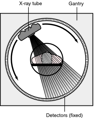

Computed tomography. Relative position of the x-ray tube, patient, and detectors in a fourth generation CT unit.
electron beam computed tomography (EBCT) ultrafast computed tomography done with a scanner in which the patient is surrounded by a large circular anode that emits x-rays as the electron beam is guided around it.
extended narrow tomography tomography involving an increase in amplitude and increase in exposure angle resulting in greater thinness of the cut for examination.
linear tomography tomography in which the tube and film move in the same direction.
narrow angle tomography a type of tomography that results in thicker sections for examination.
pluridirectional tomography tomography in which there is a great deal of movement in a variety of directions.
positron emission tomography (PET) a combination of computed tomography and scintillation scanning. Natural biochemical substances or drugs tagged with a positron-emitting radioisotope are administered to the subject by injection; the tagged substance (tracer) then becomes localized in specific tissues like its natural analogue. When the isotope decays, it emits a positron, which then annihilates with an electron of a nearby atom, producing two 511 keV gamma rays traveling in opposite directions 180 degrees apart. When the gamma rays trigger a ring of detectors around the subject, the line between the detectors on which the decay occurred is stored in the computer. A computer program (reconstruction algorithm), like those used in computed tomography, produces an image of the distribution of the tracer in the plane of the detector ring.
Most of the isotopes used in PET scanning have a half-life of only 2 to 10 minutes. Therefore, they must be produced by an on-site cyclotron and attached chemically to the tracer and used within minutes. Because of the expense of the scanner and cyclotron, PET is used only in research centers. However, PET is important because it provides information that cannot be obtained by other means. By labeling the blood with 11C-carbon monoxide, which binds to hemoglobin, images can be obtained showing the regional perfusion of an organ in multiple planes. By using labeled metabolites, images can be obtained showing metabolic activity of an organ. 15O-oxygen and 11C-glucose have been used for brain imaging and 11C-palmitate for heart imaging. 81Rb, which is distributed like potassium, is also used for heart imaging. By using labeled neurotransmitters, hormones, and drugs the distribution of receptors for these substances in the brain and other organs can be mapped.
Most of the isotopes used in PET scanning have a half-life of only 2 to 10 minutes. Therefore, they must be produced by an on-site cyclotron and attached chemically to the tracer and used within minutes. Because of the expense of the scanner and cyclotron, PET is used only in research centers. However, PET is important because it provides information that cannot be obtained by other means. By labeling the blood with 11C-carbon monoxide, which binds to hemoglobin, images can be obtained showing the regional perfusion of an organ in multiple planes. By using labeled metabolites, images can be obtained showing metabolic activity of an organ. 15O-oxygen and 11C-glucose have been used for brain imaging and 11C-palmitate for heart imaging. 81Rb, which is distributed like potassium, is also used for heart imaging. By using labeled neurotransmitters, hormones, and drugs the distribution of receptors for these substances in the brain and other organs can be mapped.
single-photon emission computed tomography (SPECT) a type of tomography in which gamma photon–emitting radionuclides are administered to patients and then detected by one or more gamma cameras rotated around the patient. From the series of two-dimensional images produced, a three-dimensional image can be created by computer reconstruction. The technique improves resolution of, and decreases interference by, overlapping organs. It is used particularly for assessment of cardiac disease, stroke, and liver disease; for staging of cancer; and to diagnose physical abnormalities through evaluation of function.
ultrasonic tomography the ultrasonographic visualization of a cross-section of a predetermined plane of the body; see B-mode ultrasonography.
Miller-Keane Encyclopedia and Dictionary of Medicine, Nursing, and Allied Health, Seventh Edition. © 2003 by Saunders, an imprint of Elsevier, Inc. All rights reserved.
to·mog·ra·phy
(tō-mog'ră-fē),Making of a radiographic image of a selected plane by means of reciprocal linear or curved motion of the x-ray tube and film cassette; images of all other planes are blurred ("out of focus") by being relatively displaced on the film.
Synonym(s): conventional tomography, planigraphy, planography, sectional radiography, stratigraphy
Farlex Partner Medical Dictionary © Farlex 2012
to·mog·ra·phy
(tŏ-mog'ră-fē)Making a radiographic image of a selected plane by means of reciprocal linear or curved motion of the x-ray tube and film cassette; images of all other planes are blurred ("out of focus") by being relatively displaced on the film.
Synonym(s): planigraphy, planography, stratigraphy.
Synonym(s): planigraphy, planography, stratigraphy.
Medical Dictionary for the Health Professions and Nursing © Farlex 2012
stratigraphy
the science of rock strata often interpreted as the study of historical geology; it is concerned with the original succession, age relations (often through fossils), and the form, distribution and composition of the rocks.Collins Dictionary of Biology, 3rd ed. © W. G. Hale, V. A. Saunders, J. P. Margham 2005
to·mog·ra·phy
(tŏ-mog'ră-fē)Making a radiographic image of a selected plane by means of reciprocal linear or curved motion of the x-ray tube and film cassette; images of all other planes are blurred ("out of focus") by being relatively displaced on the film.
Synonym(s): planigraphy, planography, stratigraphy.
Synonym(s): planigraphy, planography, stratigraphy.
Medical Dictionary for the Dental Professions © Farlex 2012
Latest Searches:
zygomaticofrontal - zygomaticofacialis - zygomaticofacial - zygomaticoauricular - zygomatico - zygomatici - zygomatica - zygomatic - zygoma - zygomas - zygodactyly - Zygocotyle - zygion - zygia - zygapophysis - zygapophysiales - zygapophysial - zygapophyseales - zygapophyseal - zygal -
- Service manuals - MBI Corp