Medical term:
Pulmo
pulmo-
, pulmon-pulmono-The lungs.
See also: pneum-, pneumo-.
See also: pneum-, pneumo-.
[L. pulmo, lung]
Farlex Partner Medical Dictionary © Farlex 2012
pulmo-
, pulmon- , pulmono-Combining forms meaning the lungs.
See also: pneum-, pneumo-
See also: pneum-, pneumo-
[L. pulmo, lung]
Medical Dictionary for the Health Professions and Nursing © Farlex 2012
lung
[lung]either of two large organs lying within the chest cavity on either side of the heart; they supply the blood with oxygen inhaled from the outside air and dispose of waste carbon dioxide in the exhaled air, as a part of the process known as respiration. Other functions include filtration of blood, serving as reservoirs to store blood, and playing a role in metabolic activities. See also color plates.
The lungs are made of elastic tissue filled with interlacing networks of tubes and sacs carrying air, and with blood vessels carrying blood. The bronchi, which bring air to the lungs, branch out within the lungs into many smaller tubes, the bronchioles, which culminate in clusters of tiny air sacs called alveoli, whose total runs into millions. The alveoli are surrounded by a network of capillaries. Through the thin membranes of the capillaries, the air and blood make their exchange of oxygen and carbon dioxide.
The lungs are divided into lobes, the left lung having two (the left upper lobe and the left lower lobe) and the right having three (the right upper lobe, the right middle lobe, and the right lower lobe); these are further subdivided into bronchopulmonary segments, of which there are about 20. Protecting each lung is the pleura, a two-layered membrane that envelops the lung and contains lubricating fluid between its inner and outer layers.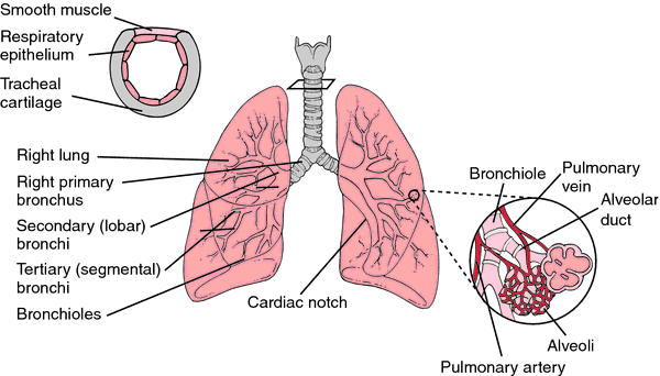
Mechanics of Inflation and Deflation. The lungs are inflated by action of the diaphragm and the intercostal muscles. The diaphragm, a large dome-shaped muscle, forms the bottom of the thoracic cage. As it contracts it flattens, increasing the diameter of the thorax and elevating the lower ribs. Both of these actions increase the space for expansion of the lungs. The external intercostal muscles provide flexibility to the thoracic cage and allow more room for lung expansion by elevating the anterior end of each rib, thereby increasing the anterior-posterior diameter of the chest wall.
Deflation of the lungs is chiefly a passive maneuver. The major muscles involved in exhalation are the abdominal muscle group. As these muscles contract, they depress the lower ribs, and, through an increase in abdominal pressure, move the diaphragm upward.
As the lungs are compressed and distended by the respiratory muscles, the pressure within the alveoli (intra-alveolar pressure) rises and falls. During inhalation the pressure becomes slightly negative (−3 mm Hg) in relation to atmospheric pressure. During exhalation the intra-alveolar pressure rises to approximately +3 mm Hg. The effect of negative pressure within the alveoli is to cause air under atmospheric pressure to flow into the lungs (inhalation). The condition of positive pressure creates the opposite effect, causing air to flow outward (exhalation).
The lungs are surrounded by an airtight compartment, the pleural space within the pleural membrane. The intrapleural pressure is less than atmospheric pressure and is expressed as negative pressure. Normally the intrapleural pressure is about −4 mm Hg. When the lungs are fully expanded this pressure may be as great as −9 mm Hg. Under normal conditions, however, the intrapleural pressure fluctuates between −4 and −6 mm Hg.
If anything should penetrate the walls of the pleura, the negative pressure is lost as air rushes into the pleural cavity in response to atmospheric pressure. This condition is called pneumothorax. The walls of the alveoli also must remain intact in order to maintain normal intrapleural pressure. If a lesion causes a break in the alveolar membranes, air enters the pleural cavity through the break and produces pneumothorax. Relief of pneumothorax and collapse of the lung from accumulations of either air or fluids within the pleural space may be provided by aspiration of the air or fluid from the thoracic cavity (thoracentesis) or by insertion of chest tubes to provide for a gradual reexpansion of the lung. (Specific tests to determine pulmonary volume and capacities are discussed under pulmonary function tests.)
Disorders of the Lungs. The air brought to the lungs is filtered, moistened, and warmed on its way along the respiratory tract but it can nevertheless bring irritants and infectious organisms, and when the body resistance is low for any reason the lungs may suffer diseases of some seriousness. Such diseases include tuberculosis and pneumonia. Other disorders of the lungs include pulmonary edema, pleurisy, asthma, bronchiectasis, atelectasis, emphysema, and pneumoconiosis. Still other diseases enter the lungs via pathogens in the circulation, and the lungs may also be affected by pulmonary embolism and chronic obstructive pulmonary disease.

Structure of the lung. From Applegate, 2000.
lung abscess an infection of the lung, characterized by a localized accumulation of pus and destruction of tissue. It may be a complication of pneumonia or tuberculosis. A lung abscess may also follow a period of excessive drinking by an alcoholic. Infected matter that has been aspirated (usually in a drunken stupor) may lodge in a bronchiole and produce inflammation. Lung cancer may also be responsible for formation of an abscess.
The first symptoms include a dry cough and chest pain. Later these may be followed by fever, chills, productive cough, headache, perspiration, foul-smelling sputum, and sometimes dyspnea. If the abscess is a complication of pneumonia, the symptoms tend to be moderated to an exaggeration of the pneumonia symptoms.
When a lung abscess forms, it is in the acute stage and treatment with antibiotics usually is effective. postural drainage may be prescribed to assist in drainage of exudate from lungs and bronchioles. In most cases, this treatment produces a cure. If the abscess becomes chronic, surgery may be necessary and usually involves removal of the portion of the lung containing the abscess.
accessory lung pulmonary sequestration.
bird breeder's lung pigeon breeder's lung.
black lung coal workers' pneumoconiosis.
brown lung byssinosis.
lung cancer malignant growths of the lung. Although the exact cause of lung cancer is not known, inhaled carcinogens are known to be important predisposing causes. Cancer in the lungs may also be a metastasis of malignancy elsewhere in the body. Many years ago it was realized that miners of certain ores who inhaled the mine dust developed lung cancer much more often than workers in other occupations. Later, other carcinogens of lung tissue, such as air polluted by fumes from burning fuels or motor exhausts, were singled out as probable causes of the increasing number of cases of the disease in urban and industrial areas. The most obvious carcinogen, however, and the one most widely encountered, is tobacco smoke, especially cigarette smoke, which is much more frequently and deeply inhaled than the smoke of pipes or cigars.
A study based on autopsies of the lungs of individuals who had died from many varied causes, but whose smoking history was known, showed that unrecognized cancer and precancerous changes in tissue were numerous among smokers and rare among nonsmokers. These findings led the Surgeon General of the United States to appoint an investigative committee, which ultimately issued a report stating that “cigarette smoking is a health hazard of sufficient importance in the United States to warrant appropriate action.”
Since the factors causing lung cancer act slowly and may produce a tumor near the periphery of the lung, early symptoms are vague or may not appear at all, and nearly a third of the cases are in an advanced stage when they are discovered. The earliest and most common symptom is a cough. Dry at first, this cough later produces sputum, which eventually becomes blood-streaked. An isolated persistent wheeze in the chest is frequently a symptom and indicates a partial obstruction in a bronchus. Chest pains, weakness, and loss of weight are later symptoms, as is dyspnea.
Diagnosis depends on a careful physical examination, including a chest x-ray. If a suspicious density is seen on the x-ray, samples of sputum will be examined microscopically for the presence of malignant cells. bronchoscopy is also done, and at the same time a specimen for biopsy can be obtained or the bronchial secretions can be washed out and the cells stained and examined.
When examination indicates lung cancer, prompt treatment is essential. This may involve the surgical removal of the lobe of the lung containing the cancer or of an entire lung if the malignant cells have spread. A significant number of persons affected by lung cancer can be cured by such operations if the surgery is performed in time. In some cases of widespread involvement surgery is not possible; these patients are treated with radiation therapy and antineoplastic drugs.
Carcinogens that can trigger lung cancer must be avoided and, when possible, eliminated. Mine workers should take adequate precautions to avoid inhaling harmful dusts. Public health authorities and industry must act more effectively to control air pollution. The most important step toward protection against lung cancer is elimination of cigarette smoking. State and local units of the American Lung Association are excellent sources of information about lung disease and its prevention.
Lung cancer clinical guidelines have been published in both the United States and Canada. In Canada they are available at the web site of Cancer Care Ontario, http://www.cancercare.on.ca. and in the United States they are available at the web site of the National Guideline Clearinghouse, http://www.guideline.gov.
coal miner's lung coal workers' pneumoconiosis.
farmer's lung hypersensitivity pneumonitis caused by inhalation of moldy hay dust.
iron lung popular name for Drinker respirator.
pigeon breeder's lung hypersensitivity pneumonitis caused by inhalation of particles of bird feces, seen in those who work closely with pigeons or other birds; it may eventually result in pulmonary fibrosis.
shock lung acute respiratory distress syndrome.
wet lung
1. pulmonary edema.
2. acute respiratory distress syndrome.
Miller-Keane Encyclopedia and Dictionary of Medicine, Nursing, and Allied Health, Seventh Edition. © 2003 by Saunders, an imprint of Elsevier, Inc. All rights reserved.
lung
(lŭng), [TA]One of a pair of viscera occupying the pulmonary cavities of the thorax, the organs of respiration in which blood is aerated. In humans, the right lung is slightly larger than the left and is divided into three lobes (an upper, a middle, and a lower or basal), whereas the left has but two lobes (an upper and a lower or basal). Each lung is irregularly conic, presenting a blunt upper extremity (the apex), a concave base following the curve of the diaphragm, an outer convex surface (costal surface), a generally concave inner or medial surface (mediastinal surface), a thin and sharp anterior border, and a rounded posterior border.
Synonym(s): pulmo [TA]
[A.S. lungen]
Farlex Partner Medical Dictionary © Farlex 2012
lung
(lŭng) [TA]One of a pair of viscera occupying the pulmonary cavities of the thorax, the organs of respiration in which aeration of the blood takes place. As a rule, the right lung is slightly larger than the left and is divided into three lobes (an upper, a middle, and a lower or basal), whereas the left has but two lobes (an upper and a lower or basal). Each lung is irregularly conic, presenting a blunt upper extremity (the apex), a concave base following the curve of the diaphragm, an outer convex surface (costal surface), an inner or mediastinal surface, a thin and sharp anterior border, and a thick and rounded posterior border.
Synonym(s): pulmo [TA] .
Synonym(s): pulmo [TA] .
[A.S. lungen]
Medical Dictionary for the Health Professions and Nursing © Farlex 2012
Latest Searches:
zygomaticofrontal - zygomaticofacialis - zygomaticofacial - zygomaticoauricular - zygomatico - zygomatici - zygomatica - zygomatic - zygoma - zygomas - zygodactyly - Zygocotyle - zygion - zygia - zygapophysis - zygapophysiales - zygapophysial - zygapophyseales - zygapophyseal - zygal -
- Service manuals - MBI Corp