Medical term:
Stenosed
stenosed
[stĕ-nōzd´]ste·nosed
(ste-nozd'),stenosed
(stə-nōzd′, -nōst′)ste·nosed
(stĕ-nōst')stenosis
(ste-no'sis) [Gr. stenosis, a narrowing]Etiology
Stenosis may result from embryonic maldevelopment, hypertrophy and thickening of a sphincter muscle, inflammatory disorders, or excessive development of fibrous tissue. It may involve almost any tube or duct.
aortic stenosis
Symptoms
Many patients with mild or moderate aortic stenosis, e.g., with a valve area that is more than 1 cm2 or a valve gradient that is less than 50 mm Hg, have no symptoms and are unaware of their condition. A heart murmur is usually heard on physical examination of the patient. This murmur is best heard at the right second intercostal space during systole. Palpation of the arteries in severe aortic stenosis may reveal a delayed and weakened pulse, e.g., at the carotids. The heart's apical impulse may be laterally and inferiorly displaced as a result of left ventricular hypertrophy. Alarming symptoms include anginal chest pain, syncope, and dyspnea on exertion. When these occur, surgery to repair or replace the diseased valve are necessary.
Physical Findings
Transthoracic echocardiography (TTE) diagnoses aortic stenosis and helps to evaluate its severity, determine left ventricular size and function, and detect other valvular disease.
Treatment
If the aortic valve area is significantly narrowed, i.e., < 0.8 cm2, or if the patient has experienced symptoms of heart failure or syncope, percutaneous balloon aortic valvuloplasty or aortic valve replacement may be necessary.
Patient care
A history of related cardiac disorders is obtained. Cardiopulmonary function is assessed regularly by monitoring vital signs and weight, intake, and output for signs of fluid overload. The patient is monitored for chest pain, which may indicate cardiac ischemia, and the electrocardiogram is evaluated for ischemic changes. Activity tolerance and fatigue are assessed.
After cardiac catheterization, the insertion site is checked according to protocol (often every 15 min for 6 hr) for signs of bleeding; the patient is assessed for chest pain, and vital signs, heart rhythm, and peripheral pulses distal to the insertion site are monitored. Problems are reported to the cardiologist.
Desired outcomes for all aortic valve surgeries include adequate cardiopulmonary tissue perfusion and cardiac output, reduced fatigue with exertion, absence of fluid volume excess, and ability to manage the treatment regimen. Patients with aortic stenosis (with or without surgical repair) require prophylactic antibiotics before invasive procedures (including dental extractions, cleanings) because of the risk they pose for bacteremia and infective endocarditis.
cicatricial stenosis
coronary artery stenosis
diaphyseal medullary stenosis
Hardcastle syndrome.infantile hypertrophic pyloric stenosis
Pyloric stenosis.lumbar spinal stenosis
mitral stenosis
Abbreviation: MSThe abnormality of the valve may predispose patients to infective endocarditis; to left atrial enlargement and atrial arrhythmias; or to left ventricular failure.
pulmonary stenosis
pyloric stenosis
Treatment
In infants, treatment may involve open or laparoscopic division of the muscles of the pylorus. Infantile pyloric stenosis is usually diagnosed in the first 6 months of life when babies have trouble with vomiting after eating, sometimes with projectile vomiting and consequent dehydration. The disease occurs in 2 to 3 infants per 1000 births and is more common in boys than girls. In adults, endoscopic stents may be placed to open malignant obstructions.
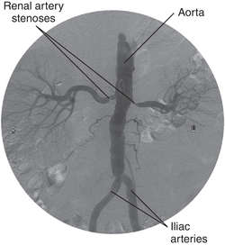
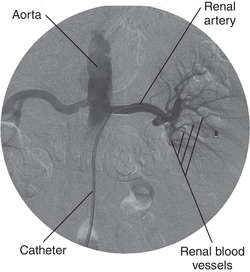
renal artery stenosis
Treatment
Patients may be treated medically with standard antihypertensive drugs, or, in some cases, with renal artery angioplasty or bypass surgery.
See: illustrationsubaortic stenosis
tricuspid stenosis
Latest Searches:
veneniferous - venenation - venenata - veneered - veneer - venectomy - venectasia - venation - Venastat - venarum - VenaFlow - venae - venacavography - venacavogram - venacaval - vena - velum - Veltin - Veltap - Velpeau -
- Service manuals - MBI Corp