Medical term:

cardioversion


Cardioversion


Definition

Cardioversion refers to the process of restoring the heart's normal rhythm by applying a controlled electric shock to the exterior of the chest.

Purpose

When the heart beats too fast, blood no longer circulates effectively in the body. Cardioversion is used to stop this abnormal beating so that the heart can begin normal rhythm and pump more efficiently.

Precautions

Not all unusual heart rhythms (called arrhythmias) are dangerous or fatal. Atrial fibrillation and atrial flutter often revert to normal rhythms without the need for cardioversion. Healthcare providers may also try to correct the heart rhythm with medication or recommend a lifestyle change before trying cardioversion. However, ventricular tachycardia lasting more than 30 seconds and ventricular fibrillation require immediate cardioversion.

Description

Elective cardioversion is usually scheduled ahead of time. After arriving at the hospital, an intravenous (IV) catheter will be placed in the arm and oxygen will be given through a face mask. A short-acting general anesthetic will be administered through the vein. During the two or three minutes of anesthesia, the doctor will apply two paddles to the exterior of the chest and administer the electric shock. It may be necessary to give the shock two or three times to obtain normal rhythm.

Preparation

Medication to thin the blood is usually given for at least three weeks before elective cardioversion. Food intake should be stopped eight hours before the procedure.

Aftercare

Medical personnel will monitor the heart rhythm for a few hours, after which the patient is usually sent home. It is advisable to arrange for transportation home, because drowsiness may last several hours. The doctor may prescribe anti-arrhythmic medication to prevent the abnormal rhythm from returning.

Risks

Cardioverters have been in use for many years and the risks are few. Those unlikely risks that remain include those instances when the device delivers greater or lesser power than expected or when power setting and control knobs are not set correctly. Unfortunately, in a number of cases, the heart prefers its abnormal rhythm and reverts to it despite cardioversion.

Normal results

Most cardioversions are successful and, at least for a time, restore the normal heart rhythm.

Key terms

Atrial fibrillation — A condition in which the upper chamber of the heart quivers instead of pumping in an organized way.
Atrial flutter — A rapid pulsation of the upper chamber of the heart that interferes with normal function.
Ventricular fibrillation — A condition in which the lower chamber of the heart quivers instead of pumping in an organized way.
Ventricular tachycardia — A rapid heart beat, usually over 100 beats per minute.

Resources

Organizations

American Heart Association. 7320 Greenville Ave. Dallas, TX 75231. (214) 373-6300. <http://www.americanheart.org>.
Gale Encyclopedia of Medicine. Copyright 2008 The Gale Group, Inc. All rights reserved.

cardioversion

 [kahr´de-o-ver″zhun]
the delivery of a direct current countershock synchronized with the QRS complex to the myocardium as an elective treatment to end tachydysrhythmias. (For emergency treatment using a nonsynchronized current to terminate arrhythmia, see defibrillation.) The goal of cardioversion is to restore sinoatrial control of the heart rhythm by depolarizing the entire myocardium at the moment of shock. The depolarization interrupts reentry circuits, thus ending myocardial fibrillation and some other types of dysrhythmias. The electric shock can be delivered directly to the myocardium in an open chest procedure, or through externally applied paddles placed on the chest.

Cardioversion is most effective in terminating arrhythmias due to continuous reentry, including atrial flutter, atrial fibrillation, paroxysmal supraventricular tachycardia, ventricular tachycardia, and ventricular fibrillation. Patients who have had a recent myocardial infarction and resultant atrial, nodal, or ventricular tachycardia are the most frequent candidates for cardioversion. Those with severe, longstanding arrhythmias due to chronic extensive heart disease usually do not benefit from this procedure.

Cardioversion should be done only by trained physicians in a setting where resuscitation equipment and respiratory support are readily at hand. Serum potassium levels must be within normal limits at the time of procedure because hypokalemia increases the patient's chance of developing deadly postconversion dysrhythmias. If necessary, potassium salts can be given prior to the procedure. digitalis toxicity predisposes the patient to life-threatening dysrhythmiasduring cardioversion and the drug should be withheld several days prior to the anticipated procedure. hypoxia and acidosis may decrease the chances of successful cardioversion.
Miller-Keane Encyclopedia and Dictionary of Medicine, Nursing, and Allied Health, Seventh Edition. © 2003 by Saunders, an imprint of Elsevier, Inc. All rights reserved.

car·di·o·ver·sion

(kar'dē-ō-ver'zhŭn),
Restoration of the heart's rhythm to normal by electrical countershock or by medications (chemical cardioversion).
[cardio- + conversion]
Farlex Partner Medical Dictionary © Farlex 2012

cardioversion

(kär′dē-ō-vûr′zhən)
n.
The restoration of the heartbeat to normal functioning by the application of electrical shock or by the use of medication.
The American Heritage® Medical Dictionary Copyright © 2007, 2004 by Houghton Mifflin Company. Published by Houghton Mifflin Company. All rights reserved.

cardioversion

Interventional cardiology The conversion of a cardiac arrhythmia, usually a tachyarrhythmia to a normal sinus rhythm; CV is most effective in terminating tachycardias due to defective reentry–eg, atrial flutter, A Fib, AV nodal entry, WPW syndrome, V tach with a pulse, flutter; the electric shock depolarizes all excitable myocardium, prolongs refractoriness, interrupts reentry circuits, and establishes electrical homogeneity; it is attempted in Pts with AFib in order to improve cardiac function, relieve symptoms, and ↓ risk of thrombus formation; transesophageal echocardiography identifies Pts with atrial emboli requiring short-term anticoagulation with heparin before cardioversion. See Chemical cardioversion, Direct current cardioversion, Single-pulse cardioversion. Cf Defibrillation.
McGraw-Hill Concise Dictionary of Modern Medicine. © 2002 by The McGraw-Hill Companies, Inc.

car·di·o·ver·sion

(kahr'dē-ō-vĕr'zhŭn)
Restoration of the heart's rhythm to normal by electrical countershock.
Medical Dictionary for the Health Professions and Nursing © Farlex 2012

cardioversion

(kar'de-o-ver?zhun) [? + L. versio, a turning]
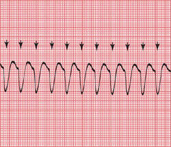
Enlarge picture
CARDIOVERSION: Synchronization during cardioversion of ventricular tachycardia; an arrow above each QRS complex denotes the period in cardiac depolarization when a shock will be delivered to terminate the rhythm
The restoration of normal sinus rhythm by chemical or electrical means. When performed medicinally, the procedure relies on the oral or intravenous administration of antiarrhythmic drugs. Electrical cardioversion relies instead on the delivery of synchronized shock of direct electrical current across the chest wall. It is used to terminate arrhythmias such as atrial fibrillation, atrial flutter, supraventricular tachycardia, and well-tolerated ventricular tachycardia. Unlike defibrillation, which is an unsynchronized shock applied during dire emergencies, electrical cardioversion is timed to avoid the T wave of cardiac repolarization to avoid triggering malignant arrhythmias. A patient will almost always require sedation and analgesia before the procedure. See: illustration

CAUTION!

Electrical cardioversion should not be used in patients who have recently eaten (because of the risk of regurgitation of stomach contents), in patients with severe electrolyte abnormalities, in patients with some drug overdoses, or in patients unable or unwilling to give informed consent. Patients need to be advised of the risks of cardioversion, including the rare precipitation of ventricular fibrillation and ventricular tachycardia, the development of bradyarrhythmias or heart blocks, and the possibility of embolic stroke.

Patient care

The procedure, expected sensations, complications, and risks are explained to and clarified for the patient. Emotional support is provided throughout the procedure and at its conclusion. The patient's medication history is reviewed, and cardiac glycoside use is reported to the health care provider, along with the patient's electrolyte levels. Emergency equipment (including ACLS drugs, a bag mask device, supplemental oxygen, suction, laryngoscope and appropriate size ET tube, defibrillator, and supplies for intravenous injection) are assembled at the bedside. In the hospital setting, emergency personnel (respiratory technicians, anesthesiologists, nurses, and paramedics) may assist the attending physician. The patient's vital signs are checked, an intravenous infusion is started, and the patient is connected to a continuous ECG monitor. Dentures are removed from the mouth, and necklaces or pendants, as well as nitroglycerin patches, are removed from the chest and neck. Chest electrodes are placed to facilitate recording of tall R waves without interfering with paddle placement. A 12-lead ECG is obtained and the patient is given enriched oxygen to breathe. The patient is placed in a supine position, and adequate ventilation and oxygenation are ensured by observation and oximetry. A sedative, such as diazepam, is provided as prescribed unless the patient is profoundly hypotensive. The defibrillator leads are attached to the patient. The cardioverter/defibrillator is set to synchronize with the patient's QRS complex, and the recording is checked to ensure that each R wave is marked. The control is set to the energy level prescribed by the health care provider or by protocol. The defibrillation pads for hands-free operation (or manual paddles) are placed in prescribed positions on the chest wall. All personnel in attendance are cleared from direct contact with the patient or his or her bed. After this is carefully verified, the electrical current is discharged. The monitor is immediately analyzed to ensure that the dysrhythmia has resolved. If it has not, the procedure is repeated, usually with a higher energy setting. After successful cardioversion, health care personnel monitor the posttreatment rhythm and vital signs until the patient's stability is assured. The patient's skin is inspected for burns. See: defibrillation

Medical Dictionary, © 2009 Farlex and Partners

cardioversion

The convertion of a dangerously rapid or irregular heart beat to normal rhythm with an electric shock. A common use is in defibrillation for one of the forms of CARDIAC ARREST or in the correction of ATRIAL FIBRILLATION.
Collins Dictionary of Medicine © Robert M. Youngson 2004, 2005

Patient discussion about cardioversion

Q. Does anyone know if its possible to get breast implants if i have an implanted defibrillator? I have hypertrophic cardiomyopathy. I'm 27 and in great health, I workout 6 days a week and I have no further symptoms.

A. Thanks for your help. In fact I have an appointment with my cardiologist in a couple of weeks and if he says its OOK I will definitely consult it with the plastic surgeon as well. However I always try to do some additional research on my own and get second opinions.
Ince again,
Thanks!

More discussions about cardioversion
This content is provided by iMedix and is subject to iMedix Terms. The Questions and Answers are not endorsed or recommended and are made available by patients, not doctors.


Latest Searches:
ZPLATE - Zovia - Zovant - Zouchlos' - Zoto - Zosyn - Zostrix - zostervirus - zosteroid - zosteriform - zoster - zorubicin - Zortress - ZORprin - Zorbtive - zorbamycin - Zorac - zopolrestat - zopiclone - Zopfius -
- Service manuals - MBI Corp