Echocardiography
Definition
Echocardiography is a diagnostic test that uses ultrasound waves to create an image of the heart muscle. Ultrasound waves that rebound or echo off the heart can show the size, shape, and movement of the heart's valves and chambers as well as the flow of blood through the heart. Echocardiography may show such abnormalities as poorly functioning heart valves or damage to the heart tissue from a past heart attack.
Purpose
Echocardiography is used to diagnose certain cardiovascular diseases. In fact, it is one of the most widely used diagnostic tests for heart disease. It can provide a wealth of helpful information, including the size and shape of the heart, its pumping strength, and the location and extent of any damage to its tissues. It is especially useful for assessing diseases of the heart valves. It not only allows doctors to evaluate the heart valves, but it can detect abnormalities in the pattern of blood flow, such as the backward flow of blood through partly closed heart valves, known as regurgitation. By assessing the motion of the heart wall, echocardiography can help detect the presence and assess the severity of coronary artery disease, as well as help determine whether any chest pain is related to heart disease. Echocardiography can also help detect hypertrophic cardiomyopathy, in which the walls of the heart thicken in an attempt to compensate for heart muscle weakness. The biggest advantage to echocardiography is that it is noninvasive (does not involve breaking the skin or entering body cavities) and has no known risks or side effects.
Precautions
Echocardiography is an extremely safe procedure and no special precautions are required.
Description
Echocardiography creates an image of the heart using ultra-high-frequency sound waves-sound waves that are too high in frequency to be heard by the human ear. The technique is very similar to ultrasound scanning commonly used to visualize the fetus during pregnancy.
An echocardiography examination generally lasts between 15-30 minutes. The patient lies bare-chested on an examination table. A special gel is spread over the chest to help the transducer make good contact and slide smoothly over the skin. The transducer, a small hand-held device at the end of a flexible cable, is placed against the chest. Essentially a modified microphone, the transducer directs ultrasound waves into the chest. Some of the waves get echoed (or reflected) back to the transducer. Since different tissues and blood all reflect ultrasound waves differently, these sound waves can be translated into a meaningful image of the heart, which can be displayed on a monitor or recorded on paper or tape. The patient does not feel the sound waves, and the entire procedure is painless. In fact, there are no known side effects.
Occasionally, variations of the echocardiography test are used. For example, Doppler echocardiography employs a special microphone that allows technicians to measure and analyze the direction and speed of blood flow through blood vessels and heart valves. This makes it especially useful for detecting and evaluating regurgitation through the heart valves. By assessing the speed of blood flow at different locations around an obstruction, it can also help to precisely locate the obstruction.
An exercise echocardiogram is an echocardiogram performed during exercise, when the heart muscle must work harder to supply blood to the body. This allows doctors to detect heart problems that might not be evident when the body is at rest and needs less blood. For patients who are unable to exercise, certain drugs can be used to mimic the effects of exercise by dilating the blood vessels and making the heart beat faster.
Preparation
The patient removes any clothing and jewelry above the chest.
Aftercare
No special measures need to be taken following echocardiography.
Risks
There are no known risks associated with the use of echocardiography.
Normal results
A normal echocardiogram shows a normal heart structure and the normal flow of blood through the heart chambers and heart valves. However, a normal echocardiogram does not rule out the possibility of heart disease.
Abnormal results
An echocardiogram may show a number of abnormalities in the structure and function of the heart, such as:
- thickening of the wall of the heart muscle (especially the left ventricle)
- abnormal motion of the heart muscle
- blood leaking backward through the heart valves (regurgitation)
- decreased blood flow through a heart valve (stenosis)
Resources
Organizations
American Heart Association. 7320 Greenville Ave. Dallas, TX 75231. (214) 373-6300. http://www.americanheart.org.
National Heart, Lung and Blood Institute. PO Box 30105, Bethesda, MD 20824-0105. (301) 251-1222. http://www.nhlbi.nih.gov.
Key terms
Noninvasive — Pertaining to a diagnostic procedure or treatment that does not require the skin to be broken or a body cavity to be entered.
Regurgitation — Backward flow of blood through a partly closed heart valve.
Transducer — A device that converts electrical signals into ultrasound waves and ultrasound waves back into electrical impulses.
Ultrasound — Sound waves at a frequency of over 20,000 kHz, often used for diagnostic imaging.
Gale Encyclopedia of Medicine. Copyright 2008 The Gale Group, Inc. All rights reserved.
ech·o·car·di·og·ra·phy
(ek'ō-kar'dē-og'ră-fē), The use of ultrasound in the investigation of the heart and great vessels and diagnosis of cardiovascular lesions.
Synonym(s): ultrasonic cardiography, ultrasound cardiography
[echo + cardiography]
Farlex Partner Medical Dictionary © Farlex 2012
echocardiography
(ĕk′ō-kär′dē-ŏg′rə-fē)n. The use of ultrasound to record and produce a two-dimensional real-time display of the size, motion, and structure of various components of the heart.
ech′o·car′di·o·graph′ic adj.
The American Heritage® Medical Dictionary Copyright © 2007, 2004 by Houghton Mifflin Company. Published by Houghton Mifflin Company. All rights reserved.
echocardiography
Doppler ultrasonography Cardiology A group of noninvasive 2-D imaging techniques that take advantage of the Doppler effect to provide information on the size, shape, and motion of cardiac structures, pressure differences in chambers, and blood flow through the heart and great vessels Applications Assess pericardial effusion, endocarditis, cardiac hypertrophy, congenital, valvular disease–mitral valve stenosis or prolapse, aortic insufficiency, aortic stenosis, subaortic stenosis, tricuspid valve, ischemic disease. See Biplane transesophageal echocardiography, Contrast echocardiography, Interventional cardiology, Intracardiac echocardiography, M-mode echocardiography, Myocardial contrast echocardiography, Stress echocardiography, Transthoracic echocardiography, Two-dimensional echocardiography. McGraw-Hill Concise Dictionary of Modern Medicine. © 2002 by The McGraw-Hill Companies, Inc.
ech·o·car·di·og·ra·phy
(ek'ō-kahr-dē-og'ră-fē) The use of ultrasound in the investigation of the structure and motion of the heart and great vessels and diagnosis of cardiovascular lesions.
Synonym(s): ultrasound cardiography.
[echo + cardiography]
Medical Dictionary for the Health Professions and Nursing © Farlex 2012
echocardiography
(ek?o-kard?e-og'ra-fe) [ echo + cardiography ] A noninvasive test that uses ultrasound to visualize cardiac structures. The heart's chambers, ejection fraction, valves, and wall motion can be evaluated, and intracardiac masses or clots can often be seen. echocardiographic (e-o-graf'ik), adjective
dobutamine stress echocardiography
Abbreviation: DSE
Echocardiography for coronary artery disease in which dobutamine is given to patients to increase the workload of the heart, and then the heart is evaluated with ultrasonic imaging. Regions of the heart that do not receive adequate blood flow (ischemic regions) contract poorly during the stress of the test but normally when the patient is at rest. Heart muscle that does not contract normally either at rest or with stimulation has been injured previously by myocardial infarction.
Doppler echocardiography
See: Doppler echocardiographymultidimensional visualization echocardiography
An experimental echocardiographic technique using computer technology for three-dimensional visualization of cardiac structures. This becomes four-dimensional when time is used to impart the cinematic perception of motion.
stress echocardiography
Echocardiography of segments of heart muscle that do not move properly when a patient with coronary artery disease exercises or takes a vasodilating drug (e.g., adenosine or dipyridamole). Stress-induced impairments in regional heart muscle activity are used as markers of obstructions in specific coronary arteries.
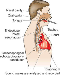

TRANSESOPHAGEAL ECHOCARDIOGRAPHY
transesophageal echocardiography
Abbreviation: TEE
A technique for obtaining echocardiographic images in which the ultrasonographic transducer is introduced into the esophagus. TEE is useful in detecting cardiac sources of emboli, prosthetic heart valve malfunction, endocarditis, aortic dissection, cardiac tumors, and valvular and congenital heart disease. See: illustration
transthoracic echocardiography
Abbreviation: TTE
Echocardiography of the heart and great vessels by means of an ultrasonic transducer placed on the chest wall. Sound waves are transmitted to the underlying organs (e.g., the heart or aorta), and an image is constructed from those that rebound toward the transducer.
Medical Dictionary, © 2009 Farlex and Partners
echocardiography
A form of ULTRASOUND imaging used to investigate heart disorders. The method can display the movement and action of the heart valves, abnormal masses in the heart and the details of congenital heart disease.Collins Dictionary of Medicine © Robert M. Youngson 2004, 2005
ech·o·car·di·og·ra·phy
(ek'ō-kahr-dē-og'ră-fē) The use of ultrasound in the investigation of the heart and great vessels and diagnosis of cardiovascular lesions.
[echo + cardiography]
Medical Dictionary for the Dental Professions © Farlex 2012
Patient discussion about echocardiography
Q. My mother had a chest pain and she was sent for a TEE. When do you need a TEE and when a normal echo is fine? My mother had a chest pain few weeks ago. we were sure its a heart attack and went to the ER. There the doctors did some tests and she was sent for a (trans thoracic echocardiogram) TEE. I want to know when do you need a TEE and when you can do just a normal echocardiogram because the TEE was very painful for her and we want to know if ther was a better way.
A. The main difference between TEE and normal echo is that in TEE u put the transducer directly in the esophagus. The transducer is the same and the idea is to put it as close as possible to the heart.
As far as I know there are some heart situations the TEE is better for diagnosis that normal echo. Maybe your mom had one of those situations?
I can recommend you to ask the ER doctor. he will probably be able to give a better explanation for his choice
Q. I am scheduled for a TEE and i am very scared. what is exactly going to happen there? I am scheduled for a Transesophageal echocardiogram (TEE) for my heart valve problem and i am very scared. what is exactly going to happen there to me? I understand i need to swallow somthing and I am not sure I'll be able to do it. I have a strong gag reflex. can someone tell me what I can do to reduce the fear?
A. During a TEE you will be requested to swallow something that looks like a big chocolate kiss. This is all the swallowing that is involved. It is not fun, and me too have a strong gag reflex. I asked the doctor to let me watch the TEE of the guy that was before me in the line. After I saw how this test is done it was easier for me.
(Don't get me wrong, you will want to puke but you will be able to handle this urge)
More discussions about echocardiographyThis content is provided by iMedix and is subject to iMedix Terms. The Questions and Answers are not endorsed or recommended and are made available by patients, not doctors.
