Medical term:
larynx
larynx
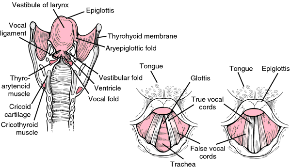
[lar´ingks] (Gr.)The larynx is composed of nine cartilages that are held together by muscles and ligament: the single thyroid, cricoid, and epiglottic cartilages and the paired arytenoid, corniculate, and cuneiform cartilages. (See also color plates.) The largest of these, the thyroid cartilage, forms the Adam's apple, which protrudes in the front of the neck. Two flexible vocal cords reach from the back to the front wall of the larynx and are manipulated by small muscles to produce sound. The epiglottis, a flap or lid at the base of the tongue, closes the larynx as it is lifted up during swallowing and so prevents passage of food or drink into the larynx and trachea.

lar·ynx
, pl.la·ryn·ges
(lar'ingks, lă-rin'jēz), Avoid the misspelling/mispronunciation larnyx.larynx
(lăr′ĭngks)larynx
The region of the throat between the pharynx (tip of the epiglottis) and trachea (cricoid cartilage) which contains the vocal cords and is involved in breathing, swallowing, and speech.Boundaries
• Superolateral boundary—Tip of the epiglottis and aryepiglottic folds.
• Inferior limit—Inferior rim of the cricoid cartilage.
• Posterior limit—Posterior mucosa covering cricoid cartilage, arytenoid region, and interarytenoid space.
• Anterior limit—Lingual surface of epiglottis, thyrohyoid membrane, anterior commissure, thyroid cartilage, cricothyroid membrane, and anterior arch of the cricoid cartilage.
Regions
Supraglottis, glottis, subglottis.
lar·ynx
, pl. larynges (laringks, lă-rinjēz) [TA]larynx
(lar'inks) plural.larynges [Gr.]
Anatomy
The framework of the larynx is built of three single cartilages and three paired cartilages. The unpaired cartilages are: the cricoid cartilage, a thick cartilage ring on top of the trachea; the thyroid cartilage, a V-shaped cartilage that sits on the cricoid with the point of its 'V' facing forward; and above this, the epiglottic cartilage, shaped like an upright paddle, with its handle held inside the front angle of the thyroid cartilage. The three smaller paired cartilages are: the arytenoids, the corniculates, and the cuneiforms. These nine cartilages are held together by membranes and ligaments, usually named by the structures that are interconnected; for example, the cricothyroid membrane connects the front of the cricoid cartilage with the base of the thyroid cartilage in the midline.
The intrinsic muscles of the larynx -- cricothyroid, posterior cricoarytenoid, lateral cricoarytenoid, thyroarytenoid, transverse and oblique arytenoids, and vocalis -- alter the length and tension of the vocal cords and the size and shape of the opening between them (the rima glottis). The vagus nerve supplies motor and sensory innervation to the larynx; the cricothyroid muscle is innervated by the external laryngeal branch of the vagus, while the other intrinsic muscles are innervated by the recurrent laryngeal branch of the vagus.
The cavity within the larynx comprises three consecutive chambers. The first chamber, the vestibule of the larynx, is a tube between the pharynx and a pair of folds, the vestibular folds (the "false vocal cords"), that protrude into the larynx. The second chamber, the ventricle of the larynx, is a short segment between the vestibular folds and the vocal folds; the ventricle has lateral recesses extending laterally under the vestibular folds. The third chamber, the infraglottic cavity (infraglottic larynx, subglottic space), is a tube between the vocal folds and the trachea.
foreign bodies in larynx
Symptoms
Symptoms may include coughing, choking, dyspnea, fixed pain, or loss of voice.
Patient care
If the patient is able to speak or cough, the rescuer should not interfere with the patient's attempts to expel the object. If the patient is unable to speak, cough, or breathe, the rescuer should apply the Heimlich maneuver 6 to 10 times rapidly in succession. Using air already in the lungs, the thrusts create an artificial cough to propel the obstructing object out of the airway. If the patient loses consciousness, carefully assist him or her to the ground in a supine (face up) position. Next the rescuer should begin CPR since compressions have been shown to be effective in clearing an obstruction. With each time attempt to ventilate, the rescuer should first look in the mouth to see if there is an object that can be pulled out of the airway with gloved fingers. Previously chest thrusts were taught for an obese or pregnant patient or a child with a foreign body airway obstruction. To simplify this procedure the Emergency Cardiac Care Guidelines 2005 recommend all patients receive chest compressions following CPR. For an infant, the rescuer uses back slaps before chest thrusts. Direct laryngoscopy and the use of Magill forceps may be required to remove a foreign object. If the object cannot be readily removed with these measures, an emergency cricothyrotomy, or emergency tracheotomy may be required. See: Heimlich maneuver
larynx
The ‘Adam's apple’ or voice box. The larynx is situated at the upper end of the wind-pipe (TRACHEA), just in front of the start of the gullet (OESOPHAGUS). At its inlet is a leaf-shaped flap of cartilage, the EPIGLOTTIS, that prevents entry of swallowed food. It has walls of cartilage and is lined with a moist mucous membrane and contains the vocal cords. These are two folds of the mucous membrane that can be tensed by tiny muscles to control their rate of vibration as air passes through them, and hence the pitch of the voice. The gap between the folds is called the glottis.larynx
a dilation of the upper part of the TRACHEA of TETRAPODS (Adam's apple in humans), occurring in the front part of the neck. It is triangular in shape (base uppermost) and is made up of 9 cartilages moved by muscles. It contains the vocal cords which are elastic ligaments embedded in two folds of mucous membrane.Larynx
lar·ynx
, pl. larynges (laringks, lă-rinjēz) [TA]larynx
[lar´ingks] (Gr.)The larynx is composed of nine cartilages that are held together by muscles and ligament: the single thyroid, cricoid, and epiglottic cartilages and the paired arytenoid, corniculate, and cuneiform cartilages. (See also color plates.) The largest of these, the thyroid cartilage, forms the Adam's apple, which protrudes in the front of the neck. Two flexible vocal cords reach from the back to the front wall of the larynx and are manipulated by small muscles to produce sound. The epiglottis, a flap or lid at the base of the tongue, closes the larynx as it is lifted up during swallowing and so prevents passage of food or drink into the larynx and trachea.

lar·ynx
, pl.la·ryn·ges
(lar'ingks, lă-rin'jēz), Avoid the misspelling/mispronunciation larnyx.larynx
(lăr′ĭngks)larynx
The region of the throat between the pharynx (tip of the epiglottis) and trachea (cricoid cartilage) which contains the vocal cords and is involved in breathing, swallowing, and speech.Boundaries
• Superolateral boundary—Tip of the epiglottis and aryepiglottic folds.
• Inferior limit—Inferior rim of the cricoid cartilage.
• Posterior limit—Posterior mucosa covering cricoid cartilage, arytenoid region, and interarytenoid space.
• Anterior limit—Lingual surface of epiglottis, thyrohyoid membrane, anterior commissure, thyroid cartilage, cricothyroid membrane, and anterior arch of the cricoid cartilage.
Regions
Supraglottis, glottis, subglottis.
lar·ynx
, pl. larynges (laringks, lă-rinjēz) [TA]larynx
(lar'inks) plural.larynges [Gr.]
Anatomy
The framework of the larynx is built of three single cartilages and three paired cartilages. The unpaired cartilages are: the cricoid cartilage, a thick cartilage ring on top of the trachea; the thyroid cartilage, a V-shaped cartilage that sits on the cricoid with the point of its 'V' facing forward; and above this, the epiglottic cartilage, shaped like an upright paddle, with its handle held inside the front angle of the thyroid cartilage. The three smaller paired cartilages are: the arytenoids, the corniculates, and the cuneiforms. These nine cartilages are held together by membranes and ligaments, usually named by the structures that are interconnected; for example, the cricothyroid membrane connects the front of the cricoid cartilage with the base of the thyroid cartilage in the midline.
The intrinsic muscles of the larynx -- cricothyroid, posterior cricoarytenoid, lateral cricoarytenoid, thyroarytenoid, transverse and oblique arytenoids, and vocalis -- alter the length and tension of the vocal cords and the size and shape of the opening between them (the rima glottis). The vagus nerve supplies motor and sensory innervation to the larynx; the cricothyroid muscle is innervated by the external laryngeal branch of the vagus, while the other intrinsic muscles are innervated by the recurrent laryngeal branch of the vagus.
The cavity within the larynx comprises three consecutive chambers. The first chamber, the vestibule of the larynx, is a tube between the pharynx and a pair of folds, the vestibular folds (the "false vocal cords"), that protrude into the larynx. The second chamber, the ventricle of the larynx, is a short segment between the vestibular folds and the vocal folds; the ventricle has lateral recesses extending laterally under the vestibular folds. The third chamber, the infraglottic cavity (infraglottic larynx, subglottic space), is a tube between the vocal folds and the trachea.
foreign bodies in larynx
Symptoms
Symptoms may include coughing, choking, dyspnea, fixed pain, or loss of voice.
Patient care
If the patient is able to speak or cough, the rescuer should not interfere with the patient's attempts to expel the object. If the patient is unable to speak, cough, or breathe, the rescuer should apply the Heimlich maneuver 6 to 10 times rapidly in succession. Using air already in the lungs, the thrusts create an artificial cough to propel the obstructing object out of the airway. If the patient loses consciousness, carefully assist him or her to the ground in a supine (face up) position. Next the rescuer should begin CPR since compressions have been shown to be effective in clearing an obstruction. With each time attempt to ventilate, the rescuer should first look in the mouth to see if there is an object that can be pulled out of the airway with gloved fingers. Previously chest thrusts were taught for an obese or pregnant patient or a child with a foreign body airway obstruction. To simplify this procedure the Emergency Cardiac Care Guidelines 2005 recommend all patients receive chest compressions following CPR. For an infant, the rescuer uses back slaps before chest thrusts. Direct laryngoscopy and the use of Magill forceps may be required to remove a foreign object. If the object cannot be readily removed with these measures, an emergency cricothyrotomy, or emergency tracheotomy may be required. See: Heimlich maneuver
larynx
The ‘Adam's apple’ or voice box. The larynx is situated at the upper end of the wind-pipe (TRACHEA), just in front of the start of the gullet (OESOPHAGUS). At its inlet is a leaf-shaped flap of cartilage, the EPIGLOTTIS, that prevents entry of swallowed food. It has walls of cartilage and is lined with a moist mucous membrane and contains the vocal cords. These are two folds of the mucous membrane that can be tensed by tiny muscles to control their rate of vibration as air passes through them, and hence the pitch of the voice. The gap between the folds is called the glottis.larynx
a dilation of the upper part of the TRACHEA of TETRAPODS (Adam's apple in humans), occurring in the front part of the neck. It is triangular in shape (base uppermost) and is made up of 9 cartilages moved by muscles. It contains the vocal cords which are elastic ligaments embedded in two folds of mucous membrane.Larynx
lar·ynx
, pl. larynges (laringks, lă-rinjēz) [TA]Latest Searches:
Voraxaze - Voranil - Voorhoeve - voodoo - VOO - Vontrol - von - vomitus - vomiturition - vomitory - vomitoria - vomito - vomitive - vomiting - vomit - vomica - vomerovaginalis - vomerovaginal - vomerorostralis - vomerorostral -
- Service manuals - MBI Corp