stenosis
[stĕ-no´sis] (pl. steno´ses) an abnormal narrowing or contraction of a body passage or opening; called also arctation, coarctation, and stricture.
aortic stenosis obstruction to the outflow of blood from the left ventricle into the aorta; in the majority of adult cases the etiology is degenerative calcific disease of the valve.
hypertrophic subaortic stenosis (idiopathic hypertrophic subaortic stenosis) a cardiomyopathy of unknown cause, in which the left ventricle is hypertrophied and the cavity is small; it is marked by obstruction to left ventricular outflow.
mitral stenosis a narrowing of the left atrioventricular orifice (mitral valve) due to inflammation and scarring; the cause is almost always rheumatic heart disease. Normally the leaflets open with each pulsation of the heart, allowing blood to flow from the left atrium into the left ventricle, and close as the ventricle fills again so that they prevent a backward flow of blood. In mitral stenosis there is a resultant increase of pressure in the pulmonary artery and hypertrophy of the left ventricle. The usual treatment is surgical replacement of the valve.
pulmonary stenosis (PS) narrowing of the opening between the pulmonary artery and the right ventricle.
pyloric stenosis see pyloric stenosis.
renal artery stenosis narrowing of one or both renal arteries by atherosclerosis or by fibrous dysplasia or hyperplasia, so that renal function is impaired (see ischemic nephropathy). Increased renin release by the affected kidney causes renovascular hypertension, and bilateral stenosis may result in chronic renal failure.
spinal stenosis narrowing of the vertebral canal, nerve root canals, or intervertebral foramina of the lumbar spine, caused by encroachment of bone upon the space; symptoms are caused by compression of the cauda equina and include pain, paresthesias, and neurogenic claudication. The condition may be either congenital or due to spinal degeneration.
subaortic stenosis aortic stenosis due to an obstructive lesion in the left ventricle below the aortic valve, causing a pressure gradient across the obstruction within the ventricle. See also idiopathic hypertrophic subaortic stenosis.
subglottic stenosis stenosis of the trachea below the glottis. A congenital form results in neonatal stridor or laryngotracheitis, often requiring tracheotomy but resolving with age. An acquired form is caused by repeated intubations.
tracheal stenosis scarring of the trachea with narrowing, usually as a result of injury from an artificial airway or trauma.
tricuspid stenosis (TS) narrowing or stricture of the tricuspid orifice of the heart, a condition often seen in patients with severe congestive heart failure, usually the result of volume overload and pulmonary hypertension with right ventricular and tricuspid annular dilation.
Miller-Keane Encyclopedia and Dictionary of Medicine, Nursing, and Allied Health, Seventh Edition. © 2003 by Saunders, an imprint of Elsevier, Inc. All rights reserved.
ste·no·sis
, pl. ste·no·ses
(ste-nō'sis, -sēz), Do not confuse this word with atresia or occlusion.A stricture of any canal or orifice.
[G. stenōsis, a narrowing]
Farlex Partner Medical Dictionary © Farlex 2012
stenosis
(stə-nō′sĭs)n. pl. steno·ses (-sēz) A constriction or narrowing of a duct or passage; a stricture.
ste·not′ic (-nŏt′ĭk) adj.
The American Heritage® Medical Dictionary Copyright © 2007, 2004 by Houghton Mifflin Company. Published by Houghton Mifflin Company. All rights reserved.
stenosis
An abnormal ↓ in the diameter of a lumen, in particular of an artery or heart valve–eg, in ASHD with plaque buildup on the inner wall of an artery. See Aortic stenosis, Carotid stenosis, Cervical stenosis, Fishmouth stenosis, Hypertrophic pyloric stenosis, Meatal stenosis, Mitral stenosis, Occlusion, Pulmonary stenosis, Restenosis, Supravalvular aortic stenosis, Subvalvular pulmonary stenosis, Supravalvular pulmonary stenosis. Cf Regurgitation. McGraw-Hill Concise Dictionary of Modern Medicine. © 2002 by The McGraw-Hill Companies, Inc.
ste·no·sis
, pl. stenoses (stĕ-nō'sis, -sēz) A stricture of any canal, especially a narrowing of one of the cardiac valves.
[G. stenōsis, a narrowing]
Medical Dictionary for the Health Professions and Nursing © Farlex 2012
stenosis
(ste-no'sis) [Gr. stenosis, a narrowing] The constriction or narrowing of a passage or orifice.
stenosedstenotic (ste-nost', ste-nozd', ) (ste-not'ik),
adjectiveEtiology
Stenosis may result from embryonic maldevelopment, hypertrophy and thickening of a sphincter muscle, inflammatory disorders, or excessive development of fibrous tissue. It may involve almost any tube or duct.
aortic stenosis
Stenosis of blood flow from the left ventricle to the aorta due to aortic valve disease or obstructions just above or below the valve. The stenosis may be congenital or secondary to diseases of adolescence or adulthood, e.g., rheumatic fever or fibrocalcific degeneration of the valve. It is the most common cardiac valve dysfunction in the U.S. Synonym: aortostenosis
Symptoms
Many patients with mild or moderate aortic stenosis, e.g., with a valve area that is more than 1 cm2 or a valve gradient that is less than 50 mm Hg, have no symptoms and are unaware of their condition. A heart murmur is usually heard on physical examination of the patient. This murmur is best heard at the right second intercostal space during systole. Palpation of the arteries in severe aortic stenosis may reveal a delayed and weakened pulse, e.g., at the carotids. The heart's apical impulse may be laterally and inferiorly displaced as a result of left ventricular hypertrophy. Alarming symptoms include anginal chest pain, syncope, and dyspnea on exertion. When these occur, surgery to repair or replace the diseased valve are necessary.
Physical Findings
Transthoracic echocardiography (TTE) diagnoses aortic stenosis and helps to evaluate its severity, determine left ventricular size and function, and detect other valvular disease.
Treatment
If the aortic valve area is significantly narrowed, i.e., < 0.8 cm2, or if the patient has experienced symptoms of heart failure or syncope, percutaneous balloon aortic valvuloplasty or aortic valve replacement may be necessary.
Patient care
A history of related cardiac disorders is obtained. Cardiopulmonary function is assessed regularly by monitoring vital signs and weight, intake, and output for signs of fluid overload. The patient is monitored for chest pain, which may indicate cardiac ischemia, and the electrocardiogram is evaluated for ischemic changes. Activity tolerance and fatigue are assessed.
After cardiac catheterization, the insertion site is checked according to protocol (often every 15 min for 6 hr) for signs of bleeding; the patient is assessed for chest pain, and vital signs, heart rhythm, and peripheral pulses distal to the insertion site are monitored. Problems are reported to the cardiologist.
Desired outcomes for all aortic valve surgeries include adequate cardiopulmonary tissue perfusion and cardiac output, reduced fatigue with exertion, absence of fluid volume excess, and ability to manage the treatment regimen. Patients with aortic stenosis (with or without surgical repair) require prophylactic antibiotics before invasive procedures (including dental extractions, cleanings) because of the risk they pose for bacteremia and infective endocarditis.
cicatricial stenosis
Stenosis due to a contracted scar.
coronary artery stenosis
A physical obstruction to the flow of blood through the epicardial arteries, usually due to atherosclerotic plaque.
diaphyseal medullary stenosis
Hardcastle syndrome.infantile hypertrophic pyloric stenosis
Pyloric stenosis.lumbar spinal stenosis
Stenosis of the spinal canal due to degenerative or traumatic changes at the level of the lumbar vertebrae. This condition causes back pain, often associated with pain that radiates into the legs, esp. when the patient is standing. Sitting often relieves the pain. The diagnosis is performed by spinal imaging, e.g., computed tomography or magnetic resonance imaging scanning. Treatments include physical therapy, braces, analgesic agents, and spinal surgery.
mitral stenosis
Abbreviation: MS
Stenosis of the mitral valve orifice with obstruction of blood flow from the left atrium to the left ventricle. In most adults, previous bouts of rheumatic carditis are responsible for the lesion. Less often, MS may be present at birth (Lutembacher's disease), or it may develop as the mitral valve calcifies during aging.
The abnormality of the valve may predispose patients to infective endocarditis; to left atrial enlargement and atrial arrhythmias; or to left ventricular failure.
pulmonary stenosis
Stenosis of the opening into the pulmonary artery from the right cardiac ventricle.
pyloric stenosis
Stenosis of the pyloric orifice. In infants, excessive thickening of the pyloric sphincter or hypertrophy and hyperplasia of the mucosa and submucosa of the pylorus are usually responsible.
Treatment
In infants, treatment may involve open or laparoscopic division of the muscles of the pylorus. Infantile pyloric stenosis is usually diagnosed in the first 6 months of life when babies have trouble with vomiting after eating, sometimes with projectile vomiting and consequent dehydration. The disease occurs in 2 to 3 infants per 1000 births and is more common in boys than girls. In adults, endoscopic stents may be placed to open malignant obstructions.
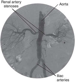

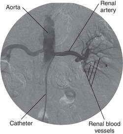
RENAL ARTERY STENOSIS: (A) Renal artery stenosis (before angioplasty); (B) Renal artery stenosis (after angioplasty) (Courtesy of Arnold Klein, M.D., Northwest Permanente, P.C.)
RENAL ARTERY STENOSIS: (A) Renal artery stenosis (before angioplasty); (B) Renal artery stenosis (after angioplasty) (Courtesy of Arnold Klein, M.D., Northwest Permanente, P.C.)
renal artery stenosis
Stenosis in one or both arteries that supply the kidneys; a relatively uncommon cause of hypertension. In young women the cause is usually fibromuscular dysplasia of one or both arteries. In older people the cause is usually atherosclerosis.
Treatment
Patients may be treated medically with standard antihypertensive drugs, or, in some cases, with renal artery angioplasty or bypass surgery.
See:
illustrationsubaortic stenosis
A congenital stenosis of the aortic tract below the aortic valves. See: hypertrophic cardiomyopathy
tricuspid stenosis
Stenosis of the opening to the tricuspid valve.
Medical Dictionary, © 2009 Farlex and Partners
stenosis
Narrowing of a duct, orifice or tubular organ such as the intestinal canal or a blood vessel.Collins Dictionary of Medicine © Robert M. Youngson 2004, 2005
Stenosis
The narrowing of an opening or passage-way in the body. In arteries, stenosis is caused by a build-up of atherosclerotic plaque, disease, or other disorder.
Mentioned in: Anesthesia, General, Balloon Valvuloplasty, Cervical Spondylosis, Congenital Heart Disease, Heart Surgery for Congenital Defects, Heart Valve Repair, Hydrocephalus, Pulmonary Valve Stenosis, Pyloric Stenosis, Spinal Stenosis, Transient Ischemic Attack, Trigger Finger, Valvular Heart Disease
Gale Encyclopedia of Medicine. Copyright 2008 The Gale Group, Inc. All rights reserved.
ste·no·sis
, pl. stenoses (stĕ-nō'sis, -sēz) Stricture of any canal or orifice.
[G. stenōsis, a narrowing]
Medical Dictionary for the Dental Professions © Farlex 2012
Patient discussion about Stenosis
Q. Why does Aortic stenosis causes an enlarged heart? My father was recently diagnosed as suffering from enlarged heart due to his Aortic stenosis. what is the connection between those to conditions? As far as I understand that aortic stenosis mean that the aortic valve is too small not too large...
A. there are several explanations for the enlargement of the heart that occurs due to Aortic stenosis. the most reasonable is that the mechanical power that the heart uses makes it bigger. it easy to see it here: http://www.marvistavet.com/assets/images/aortic_stenosis.gif
this is called Left Ventricular Hypertrophy or LVH in abbreviations.
this is a classic LVH E.C.G.
http://www.frca.co.uk/images_main/resources/ECG/ECGresource39.jpg
Q. How does alcohol affect someone who has been diagnosed with aortic valve stenosis? My brother has been diagnosed with aortic valve stenosis and also is a smoker and does drink alcohol on the weekends. He knows that he should stop smoking but what about the effects of alcohol? Does this also contribute to his stenosis?
A. Alcohol changes blood pressure and speed of the heart- that is not a good idea if you have an Aortic stenosis. Could probably makes things worst. I would avoid alcohol… but he should ask GP.
Q. what does c4-5 mild central disk bulging impinging upon cervical cord without spinal stenosis or distortion of the cord . mild righ neural foraminal narrowing from uncovertebral joint hypertropy mean
A. Well this basically means there is a very small narrowing of the cervical (your neck area) spinal canal (where the spinal cord is), however the narrowing does not cause any damage to the spinal cord, therefore probably does not cause any major symptoms involving the nerves. The c4-5 bulging part refers to the part in between the two cervical vertebras c4 and c5, in which the disc (a part in the spinal cord) is sliding a bit side-ways, but again, it does not seem to be causing any trouble.
More discussions about StenosisThis content is provided by iMedix and is subject to iMedix Terms. The Questions and Answers are not endorsed or recommended and are made available by patients, not doctors.

